

Pre-structured hydrophobic peptide β-strands: A universal amyloid trap?
- PMID: 30707943
- PMCID: PMC7094768
- DOI: 10.1016/j.abb.2019.01.032
Pre-structured hydrophobic peptide β-strands: A universal amyloid trap?
Abstract
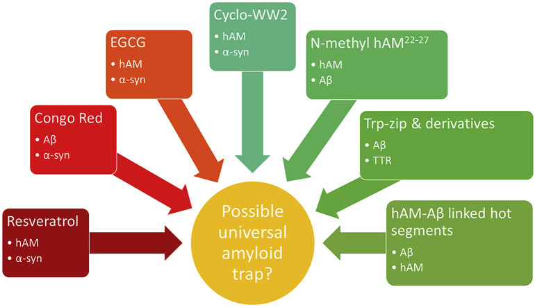
Amyloid fibril formation has long been studied because of the variety of proteins that are capable of adopting this structure despite sharing little sequence homology. This makes amyloid fibrils a challenging focus for inhibition studies because the peptides and proteins that form amyloid fibrils cannot be targeted based on a sequence motif. Most peptide inhibitors that target specific amyloidogenic proteins rely heavily on sequence recognition to ensure that the inhibitory peptide is able to bind its target. This approach is limited to targeting one amyloidogenic protein at a time. However, there is increasing evidence of cross-reactivity between amyloid-forming polypeptides. It has therefore become more useful to study the similarities between these proteins that goes beyond their sequence homology. Indeed, the observation that amyloidogenic proteins adopt similar secondary structures along the pathway to fibril formation opens the way to an interesting investigation: the development of inhibitors that could be universal amyloid traps. The review below will analyze two specific amyloidogenic proteins, α-synuclein and human amylin, and introduce a small number of peptides that have been shown to be capable of inhibiting the amyloidogenesis of both of these very dissimilar polypeptides. Some of the inhibitory peptide motifs may indeed, be applicable to Aβ and other amyloidogenic systems.
Keywords: Amylin; Amyloid; Inhibitors; Parkinson's disease; Synuclein; Type II diabetes.
Copyright © 2019 Elsevier Inc. All rights reserved.
Figures
Similar articles
-
Designed hairpin peptides interfere with amyloidogenesis pathways: fibril formation and cytotoxicity inhibition, interception of the preamyloid state.Biochemistry. 2011 Sep 27;50(38):8202-12. doi: 10.1021/bi200760h. Epub 2011 Aug 30. Biochemistry. 2011. PMID: 21848289 Free PMC article.
-
Sequence and structure-based peptides as potent amyloid inhibitors: A review.Arch Biochem Biophys. 2020 Nov 30;695:108614. doi: 10.1016/j.abb.2020.108614. Epub 2020 Sep 30. Arch Biochem Biophys. 2020. PMID: 33010227 Review.
-
Sequence-Dependent Self-Assembly and Structural Diversity of Islet Amyloid Polypeptide-Derived β-Sheet Fibrils.ACS Nano. 2017 Sep 26;11(9):8579-8589. doi: 10.1021/acsnano.7b02325. Epub 2017 Aug 3. ACS Nano. 2017. PMID: 28771324 Free PMC article.
-
Multi-Targeting Macrocyclic Peptides as Nanomolar Inhibitors of Self- and Cross-Seeded Amyloid Self-Assembly of α-Synuclein.Angew Chem Int Ed Engl. 2025 Apr 1;64(14):e202422834. doi: 10.1002/anie.202422834. Epub 2025 Jan 31. Angew Chem Int Ed Engl. 2025. PMID: 39822034
-
Elucidating the Structures of Amyloid Oligomers with Macrocyclic β-Hairpin Peptides: Insights into Alzheimer's Disease and Other Amyloid Diseases.Acc Chem Res. 2018 Mar 20;51(3):706-718. doi: 10.1021/acs.accounts.7b00554. Epub 2018 Mar 6. Acc Chem Res. 2018. PMID: 29508987 Free PMC article. Review.
Cited by
-
The hot sites of α-synuclein in amyloid fibril formation.Sci Rep. 2020 Jul 22;10(1):12175. doi: 10.1038/s41598-020-68887-2. Sci Rep. 2020. PMID: 32699326 Free PMC article.
-
Alternative Causal Link between Peptide Fibrillization and β-Strand Conformation.ACS Omega. 2021 May 5;6(19):12904-12912. doi: 10.1021/acsomega.1c01423. eCollection 2021 May 18. ACS Omega. 2021. PMID: 34056442 Free PMC article.
-
Direct Identification of Amyloid Peptide Fragments in Human α-Synuclein Based on Consecutive Hydrophobic Amino Acids.ACS Omega. 2020 May 12;5(20):11677-11686. doi: 10.1021/acsomega.0c00979. eCollection 2020 May 26. ACS Omega. 2020. PMID: 32478258 Free PMC article.
References
-
- Rochet J.C., Lansbury P.T. Amyloid fibrillogenesis: themes and variations. Curr. Opin. Struct. Biol. 2000;10:60–68. - PubMed
-
- Padrick S.B., Miranker A.D. Islet amyloid: phase partitioning and secondary nucleation are central to the mechanism of fibrillogenesis. Biochemistry. 2002;41:4694–4703. - PubMed
Publication types
MeSH terms
Substances
Grants and funding
LinkOut - more resources
Full Text Sources
