

Transcytosis at the blood-brain barrier
- PMID: 30708291
- PMCID: PMC6629499
- DOI: 10.1016/j.conb.2018.12.014
Transcytosis at the blood-brain barrier
Abstract
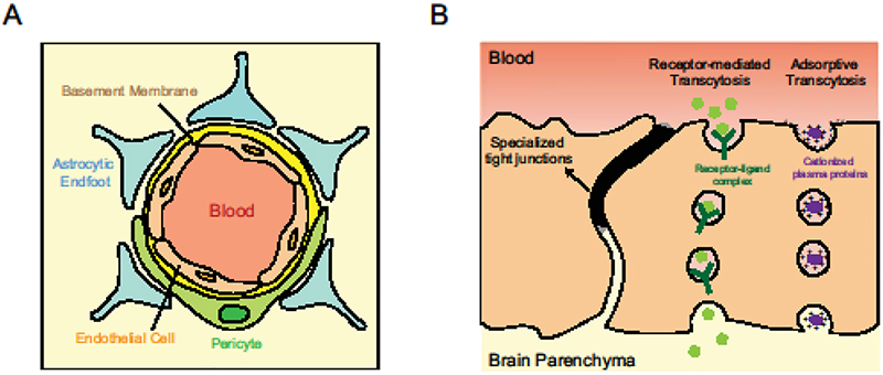
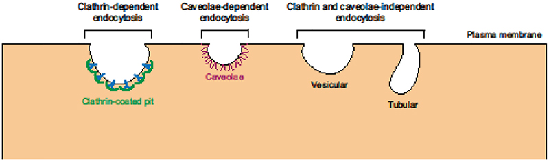
The blood-brain barrier (BBB) is a functional interface separating the brain from the circulatory system and is essential for homeostasis of the central nervous system (CNS). The BBB regulates molecular flux to maintain an optimal environment for neuronal function and protects the brain from toxins and pathogens. Endothelial cells forming the walls of CNS blood vessels constitute the BBB. CNS endothelial cells exhibit two features that underlie the restrictive properties of the BBB: specialized tight junctions that prevent paracellular passage between the blood and the brain, and unusually low levels of vesicle trafficking that limit transcellular transport or transcytosis. While the prevailing view in the field was that specialized tight junctions contributed to CNS barrier properties, recent findings have revealed the importance of maintaining low rates of transcytosis at the BBB. It is now clear that suppression of transcytosis at the BBB is an active process and CNS-specific genetic programs inhibit this pathway to maintain a functional barrier.
Copyright © 2019 Elsevier Ltd. All rights reserved.
Conflict of interest statement
Conflict of interest
The authors declare no conflict of interest.
Figures
References
-
- Potente M, Makinen T: Vascular heterogeneity and specialization in development and disease. Nat Rev Mol Cell Biol 2017, 18:477–494. - PubMed
-
- Augustin HG, Koh GY: Organotypic vasculature: From descriptive heterogeneity to functional pathophysiology. Science 2017, 357. - PubMed
-
- Tilton RG, Kilo C, Williamson JR: Pericyte-endothelial relationships in cardiac and skeletal muscle capillaries. Microvasc Res 1979, 18:325–335. - PubMed
-
- Frank RN, Dutta S, Mancini MA: Pericyte coverage is greater in the retinal than in the cerebral capillaries of the rat. Invest Ophthalmol Vis Sci 1987, 28:1086–1091. - PubMed
Publication types
MeSH terms
Grants and funding
LinkOut - more resources
Full Text Sources
Other Literature Sources
