. 2019 Jan 31;24(3):513.
doi: 10.3390/molecules24030513.

A Role of Exopolysaccharide Produced by Streptococcus thermophilus in the Intestinal Inflammation and Mucosal Barrier in Caco-2 Monolayer and Dextran Sulphate Sodium-Induced Experimental Murine Colitis

Affiliations

A Role of Exopolysaccharide Produced by Streptococcus thermophilus in the Intestinal Inflammation and Mucosal Barrier in Caco-2 Monolayer and Dextran Sulphate Sodium-Induced Experimental Murine Colitis

Yun Chen et al. Molecules. .

Abstract

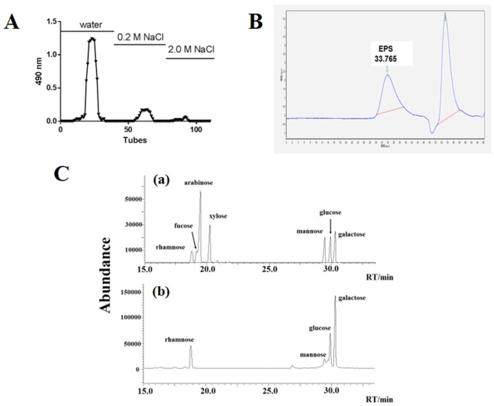
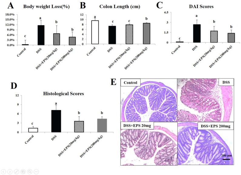
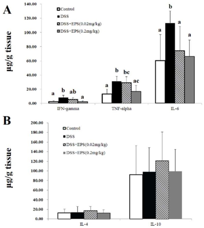
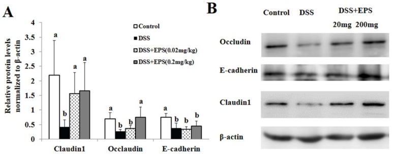
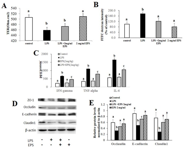
Exopolysaccharide (EPS) produced by probiotics may play an important role in gastrointestinal disease prevention, including ulcerative colitis. However, there is no literature reporting on the intervention effects of purified EPS. The aim of this study was to investigate the alleviating effect of the purified EPS produced by Streptococcus thermophilus MN-BM-A01 on murine model of colitis induced by dextran sulphate sodium (DSS). A water-soluble heteropolysaccharide (EPS-1) isolated from MN-BM-A01 was composed of rhamnose, glucose, galactose, and mannose in a molar ratio of 12.9:26.0:60.9:0.25, with molecular weight of 4.23 × 10⁵ Da. After EPS-1 administration, the disease severity of mouse colitis was significantly alleviated, mainly manifesting as the decrease of disease activity index and mitigated colonic epithelial cell injury. Meanwhile, pro-inflammatory cytokines levels (tumor necrosis factor-α, interleukin-6, and interferon-γ) were significantly suppressed, the reduced expressions of tight junction protein (claudin-1, occludin, and E-canherin) were counteracted. In addition, the results in vitro showed that EPS-1 protected intestinal barrier integrity from the disruption by lipopolysaccharide in Caco-2 monolayer, increased expression of tight junction and alleviated pro-inflammatory response. Collectively, our study confirmed the protective effects of purified EPS produced by Streptococcus thermophilus on acute colitis via alleviating intestinal inflammation and improving mucosal barrier function.

Keywords: DSS-induced colitis; Streptococcus thermophiles; exopolysaccharide; tight junction protein.


Conflict of interest statement

The authors declare no conflict of interest.

Figures

Figure 1
Figure 1
Isolation, molecular mass determination and monosaccharide composition of EPS from S. thermophilus MN-BM-A01. (A) Crude EPS separation profile by anion-exchange chromatography of DEAE Sepharose Fast flow. (B) GPC chromatogram of EPS-1. (C) Chromatogram of standard monosaccharides (a) and EPS-1 from S. thermophilus MN-BM-A01 monosaccharides (b) on chromatographic column.
Figure 2
Figure 2
EPS-1 attenuates DSS-induced acute murine colitis. (A) Body weights loss, (B) variations of colon length, (C) disease activity index (DAI), and (D) histological scores of mice from each treatment group. (E) Representative HE staining colonic tissue from each treatment group, scale bars, 200 μm. Values with different superscript letters (a, b, c, d) are significantly different (p < 0.05).
Figure 3
Figure 3
EPS-1 promoted a change in pro-inflammatory and anti-inflammatory cytokines. (A) The levels of pro-inflammatory cytokines in the colonic tissues from each treatment group. (B) The levels of anti-inflammatory cytokines in the colonic tissues from each treatment group. Values with different superscript letters (a, b, c) are significantly different (p < 0.05).
Figure 4
Figure 4
EPS-1 Relieved DSS-Induced Colonic Epithelial Tight Junction Disruption in Mice. (A) Relative protein levels of Occludin, E-cadherin and Claudin1 in the colon tissues normalized to β-actin. (B) Protein expression of Occludin, E-cadherin, and Claudin-1 in the colon tissues were analyzed by Western blot. Values with different superscript letters (a, b,) are significantly different (p < 0.05).
Figure 5
Figure 5
EPS-1 Protected intestinal barrier integrity from the disruption by LPS in Caco-2 monolayer. (A) The changes of transepithelial electrical resistance (TER) before and after treatment with LPS (10 μg/mL) and/or EPS-1 (2 mg/mL) for 24 h in Caco-2 monolayer. (B) FITC-dextran (4 kDa) permeability measurement in 21-day cultured Caco-2 monolayer after LPS and/or EPS-1 treatment. (C) The levels of pro-inflammatory cytokines in the Caco-2 cells from each treatment group. (D) Protein expression of Occludin, E-cadherin and Claudin1 in the Caco-2 cells from each treatment group. (E) Relative protein levels of Occludin, E-cadherin and Claudin1 in Caco-2 cells normalized to β-actin. Values with different superscript letters (a, b, c) are significantly different (p < 0.05).

Similar articles

Cited by

References

    1. Kaser A., Zeissig S., Blumberg R.S. Inflammatory bowel disease. Annu. Rev. Immunol. 2010;28:573–621. doi: 10.1146/annurev-immunol-030409-101225. - DOI - PMC - PubMed
    1. Lakatos P.L. Recent trends in the epidemiology of inflammatory bowel diseases: Up or down? World J. Gastroenterol. 2006;12:6102–6108. doi: 10.3748/wjg.v12.i38.6102. - DOI - PMC - PubMed
    1. Lechuga S., Ivanov A.I. Disruption of the epithelial barrier during intestinal inflammation: Quest for new molecules and mechanisms. Biochim. Biophys. Acta Mol. Cell Res. 2017;1864:1183–1194. doi: 10.1016/j.bbamcr.2017.03.007. - DOI - PMC - PubMed
    1. Al-Sadi R., Boivin M., Ma T. Mechanism of cytokine modulation of epithelial tight junction barrier. Front. Biosci. 2009;14:2765–2778. doi: 10.2741/3413. - DOI - PMC - PubMed
    1. Barreau F., Hugot J.P. Intestinal barrier dysfunction triggered by invasive bacteria. Curr. Opin. Microbiol. 2014;17:91–98. doi: 10.1016/j.mib.2013.12.003. - DOI - PubMed

MeSH terms

LinkOut - more resources