

Intraosseous synovial sarcoma of the distal ulna: a case report and review of the literature
- PMID: 30709383
- PMCID: PMC6359868
- DOI: 10.1186/s12885-019-5325-x
Intraosseous synovial sarcoma of the distal ulna: a case report and review of the literature
Abstract
Background: Synovial sarcoma is a relatively rare type of soft tissue sarcoma. The commonly observed symptom is a deep-seated palpable mass accompanied by pain or tenderness. Thus, it is considered a soft tissue sarcoma and rarely occurs primarily in bone. However, only few studies have been reported on intraosseous synovial sarcoma, and reports on cases with cytogenetic or molecular confirmation are even rarer. We report a case of intraosseous synovial sarcoma of the distal ulna that has been confirmed using histopathological examination and molecular analysis.
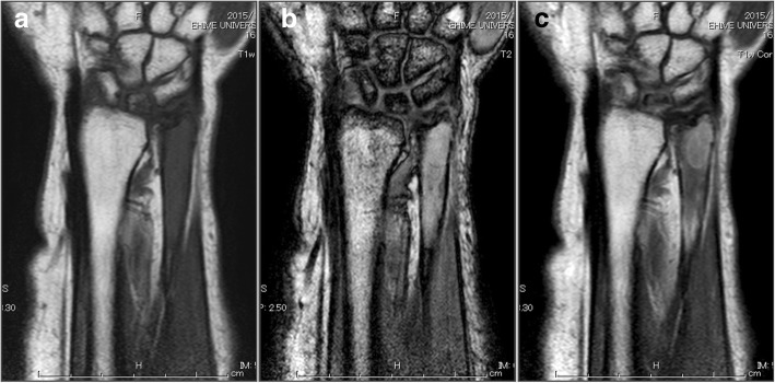
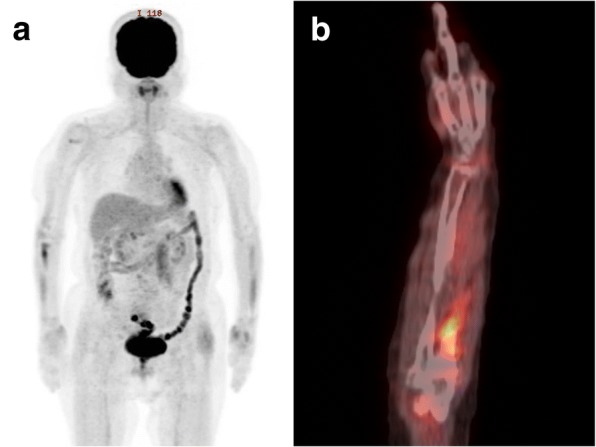
Case presentation: A 77-year-old female was referred to our hospital with a 1-month history of right wrist pain after housework. Clinical and imaging findings suggested a benign bone tumor that was enhanced by Gd-DTPA. It was thought that the tumor was possibly an enchondroma. Initially, we planned to evaluate the benignancy of the tumor with intraoperative frozen section, followed by curettage and bone graft at one stage However, when considering carefully, characteristics of the tumor did not perfectly match those of any diagnostic categories including enchondroma. Therefore, an incisional biopsy was performed and revealed that the tumor was synovial sarcoma. Following an elaborate plan, the patient underwent a wide resection of the tumor at the distal part of the right ulna. Reverse transcription-polymerase chain reaction (RT-PCR) from the resected specimen and sequencing of RT-PCR products demonstrated a chimeric SYT-SSX1 transcript, confirming the diagnosis of synovial sarcoma.
Conclusions: Synovial sarcoma is seldom considered in differential diagnosis of bone tumors because it is difficult to line up such an unusual diagnosis as a differential diagnosis. When the lesion does not perfectly fit into any diagnostic category, when the initial image diagnosis appears unconvincing, biopsy and pathology are indicated, recalling Jaffe's triangle. According to these diagnostic processes, the patient successfully completed the treatment for this rare intraosseous synovial sarcoma, following a careful plan based on the preoperative diagnosis.
Keywords: Bone tumor; SYT-SSX fusion gene; Synovial sarcoma.
Conflict of interest statement
Ethics approval and consent to participate
This report has been performed in accordance with the ethical standards in the Declaration of Helsinki. Ethics committee approval was waived since no approval is required for a case report and literature review.
Consent for publication
Written informed consent was obtained from the patient for publication of the clinical details and an individual data in any form including images, videos, voice recordings etc. A copy of the consent form is available for review by the Editor of this journal.
Competing interests
The authors declare that they have no competing interests.
Publisher’s Note
Springer Nature remains neutral with regard to jurisdictional claims in published maps and institutional affiliations.
Figures


References
-
- Goldblum JR, Weiss SW, Folpe AL. Enzinger and Weiss’s Soft Tissue Tumors. 15. 2008. Synovial sarcoma; pp. 1161–1182.
-
- Suurmeijer A, de Brujin D, Geurts van Kessel D, Miettinen M. WHO classification of Tumours of soft tissue and bone. 4. 2013. Synovial Sarcoma; pp. 213–215.
-
- Aparna M, Natarajan J, Arumugam C, Radhakrishnan R. Primary synovial sarcoma of the maxilla. J Cancer Res Ther. 2014;10:739–741. - PubMed
Publication types
MeSH terms
Substances
LinkOut - more resources
Full Text Sources
Medical
