Review
. 2019 Mar;39(1):171-183.
doi: 10.1016/j.cll.2018.10.006. Epub 2018 Dec 18.

Signaling Molecules in Posttransplantation Cancer

Affiliations
Review

Signaling Molecules in Posttransplantation Cancer

Murugabaskar Balan et al. Clin Lab Med. 2019 Mar.

Abstract

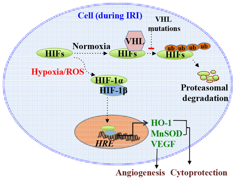
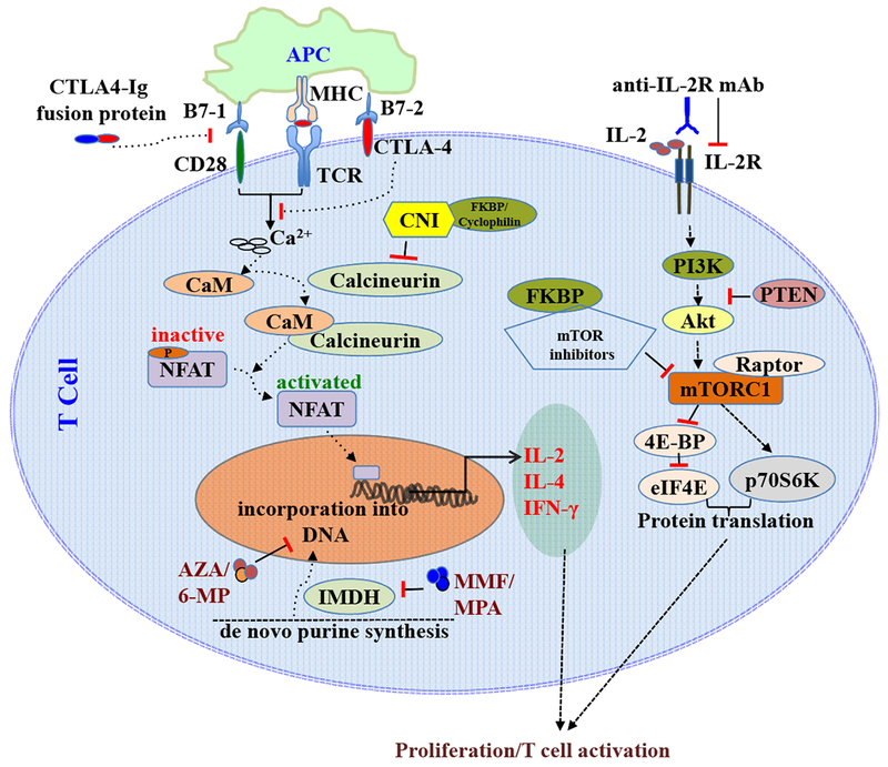
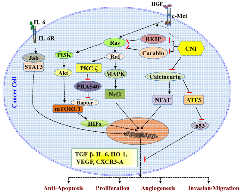
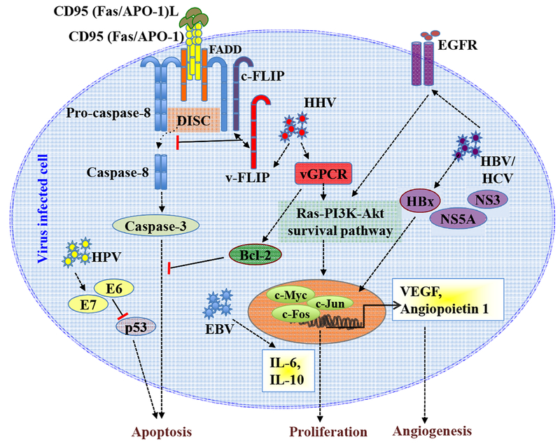
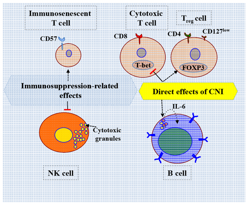
Immunosuppression is essential to prevent graft rejection. However, immunosuppression impairs the ability of the host immune system to control viral infection and decreases tumor immunosurveillance. Therefore, immunosuppression after organ transplantation is a major risk factor for posttransplantation cancer. Notably, recent reports suggest that immunosuppressive agents can activate tumorigenic pathways independent of the involvement of the host immune system. In this review, we focus on cell-intrinsic tumorigenic pathways directly activated by immunosuppressive agents and discuss the much-described infection- and immune-mediated mechanisms of cancer development in organ transplant recipients.

Keywords: CNI; Immunosuppression; Posttransplantation cancer; Risk factor; Signaling mechanisms.


Conflict of interest statement

Disclosure Statement

The authors have nothing to disclose.

Figures

FIGURE 1.
FIGURE 1.. Hypoxia-related signaling molecules in post-transplantation cancer.
Under normoxic conditions, VHL protein targets HIFs for degradation. However, during IRI, hypoxia and ROS generation activate HIFs. Activated HIFs translocate to nucleus and bind hypoxia response elements (HRE) in the promoter regions of hypoxia-inducible genes, such as HO-1 and MnSOD, and angiogenic factors (like VEGF), leading to overexpression of these molecules.
FIGURE 2.
FIGURE 2.. Molecular targets of immunosuppressive agents.
Top left: Antigen presentation via MHC on APCs and co-stimulatory interactions (like B7–1/CD28) activate TCR, and it leads to an increase in the intracellular calcium levels and activation of calmodulin (CaM), which in turn binds and activates calcineurin. Phosphorylated NFAT is inactive and resides in the cytoplasm. Activated calcineurin, with its protein phosphatase activity, dephosphorylates NFAT and promotes the nuclear translocation of NFAT to activate transcription of NFAT target genes, including IL-2, IL-4 and IFN-γ, leading to T cell activation. Co-inhibitory interactions like B7–2/CTLA-4 suppress TCR-mediated signaling. Recombinant fusion CTLA4-Ig can disrupt B7– 1/CD28 and other co-stimulatory signals necessary for T cell activation. CNIs form drug/FKBP (or cyclophilin) complex and inactivate calcineurin. Right: mTOR is a serine/threonine protein kinase and is one of the downstream effector molecules for IL-2/IL-2R (and other growth factors)-induced PI3K-Akt signaling pathways. PTEN inhibits Akt activation through PI3K suppression. mTORC1, the functional protein complex containing RAPTOR, regulates protein translation by modulating the activity of p70S6K and eIF4E (through 4E-BP). mTOR inhibitors form drug/FKBP complex and inhibit mTOR. Anti-IL-2R antibody blocks the activation of IL- 2R. Bottom left: MMF and MPA inhibit IMDH-dependent de novo synthesis of guanosine nucleoside. AZA and 6-MP antagonize endogenous purines and prevent the incorporation of guanosine into DNA.
FIGURE 3.
FIGURE 3.. Immunosuppressive agent-induced cell-intrinsic tumorigenic pathways.
Right: RTK c-Met-mediated signaling promotes the activation of Ras-Raf-MAPK pathways and the nuclear translocation of Nrf2, leading to overexpression of HO-1. CNI enhances c-Met-induced Ras activation and HO-1 expression. CNI inhibits RKIP and carabin, the negative regulators of Ras, and contributes to sustained Ras activation in cancer cells. CNI promotes the expression of ATF3, and it can inhibit p53-mediated apoptotic pathways. Center: CNI increases the expression of VEGF via PI3K-Akt-mTORC1 pathway channeled through HIFs. CNI-induced Ras-Raf-PKC-ζ activation inhibits PRAS40, the negative regulator of mTORC1, and augments mTORC1 activity. CNI treatment increases the expression of TGF-β, IL-6, and CXCR3-A (the growth promoting CXCR3 isoform). Left: IL-6-mediated signaling activates JAK-STAT3 pathway. Together, CNI-induced signaling activates cell-intrinsic pro-tumorigenic pathways.
FIGURE 4.
FIGURE 4.. Oncogenic viral infections in post-transplantation malignancies.
Top Left: Binding of CD95(Fas/APO-1)L to CD95(Fas/APO-1) initiates the formation of DISC involving pro-caspase-8 and FADD resulting in the activation of pro-caspase-8. Subsequent activation of other caspases promotes apoptosis. HHV-encoded vFLIP interferes with the activation of pro-caspase-8 at DISC and inhibits apoptosis. HHV-encoded vGPCR promotes Ras-PI3K-Akt survival pathway and increases the expression of anti-apoptotic Bcl-2. Right: HBV-encoded HBx and HCV-encoded NS3 and NS5A proteins activate Ras-PI3K-Akt survival pathway. HBx and NS5A modulate RTK EGFR-mediated signaling. HBx modulates the transcriptional activity of c-Myc, c-Fos and c-Jun and increases the expression of angiogenic factors, such as VEGF and angiopoietin-1. Bottom: HPV-encoded E6 and E7 proteins inhibit p53-mediated apoptosis. EBV infection increases the secretion of IL-6 and IL-10. Together, oncogenic virus infection disrupts the apoptotic machinery of infected cells and also promotes angiogenesis, resulting in the malignant transformation of the infected cell.
FIGURE 5.
FIGURE 5.. Immunosuppression-related impaired tumor immunosurveillance.
Important changes observed in the immune phenotype of transplant recipients, as a result of immunosuppression, are summarized. CNI regimen has been associated with an increase in T reg cells and increased production of IL-6 from B cells. Increase in immunosenescent T cells and impaired NK cell cytotoxicity were observed in organ transplant recipients treated with CNI. Also, CNI treatment downregulated T-bet expression in CD8+ T cells and drastically affected its cytotoxic effector functions in vivo. Taken together, immunosuppression alters the phenotype and functions of immune cells, leading to impaired tumor immunosurveillance.

References

    1. Au E, Wong G, Chapman JR. Cancer in kidney transplant recipients. Nat Rev Nephrol. 2018. - PubMed
    1. Sprangers B, Nair V, Launay-Vacher V, Riella LV, Jhaveri KD. Risk factors associated with post-kidney transplant malignancies: an article from the Cancer-Kidney International Network. Clin Kidney J. 2018;11(3):315–329. - PMC - PubMed
    1. van de Wetering J, Roodnat JI, Hemke AC, Hoitsma AJ, Weimar W. Patient survival after the diagnosis of cancer in renal transplant recipients: a nested case-control study. Transplantation. 2010;90(12):1542–1546. - PubMed
    1. Hall EC, Pfeiffer RM, Segev DL, Engels EA. Cumulative incidence of cancer after solid organ transplantation. Cancer. 2013;119(12):2300–2308. - PMC - PubMed
    1. Engels EA, Pfeiffer RM, Fraumeni JF, Jr., et al. Spectrum of cancer risk among US solid organ transplant recipients. JAMA. 2011;306(17):1891–1901. - PMC - PubMed

Publication types

MeSH terms