. 2019 Mar 29;294(13):4738-4758.
doi: 10.1074/jbc.RA118.005008. Epub 2019 Feb 1.

The G2019S variant of leucine-rich repeat kinase 2 (LRRK2) alters endolysosomal trafficking by impairing the function of the GTPase RAB8A

Affiliations

The G2019S variant of leucine-rich repeat kinase 2 (LRRK2) alters endolysosomal trafficking by impairing the function of the GTPase RAB8A

Pilar Rivero-Ríos et al. J Biol Chem. .

Abstract

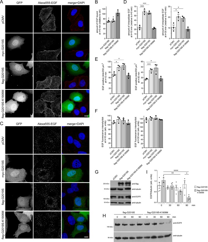
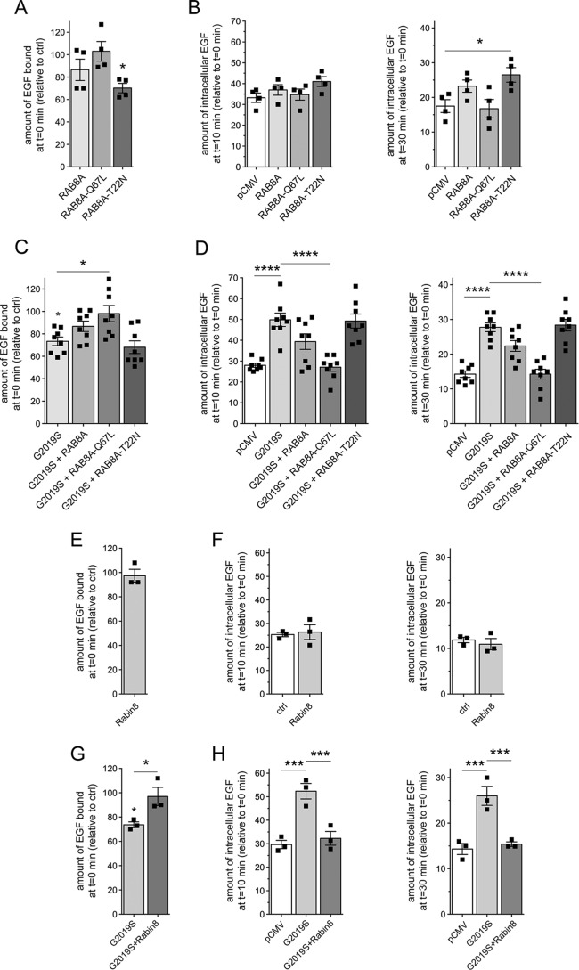
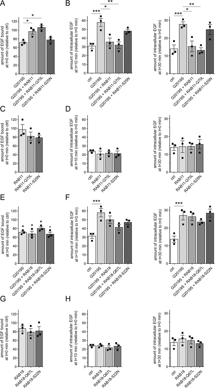
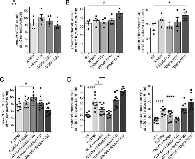
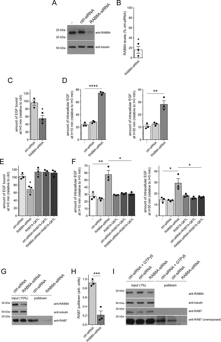
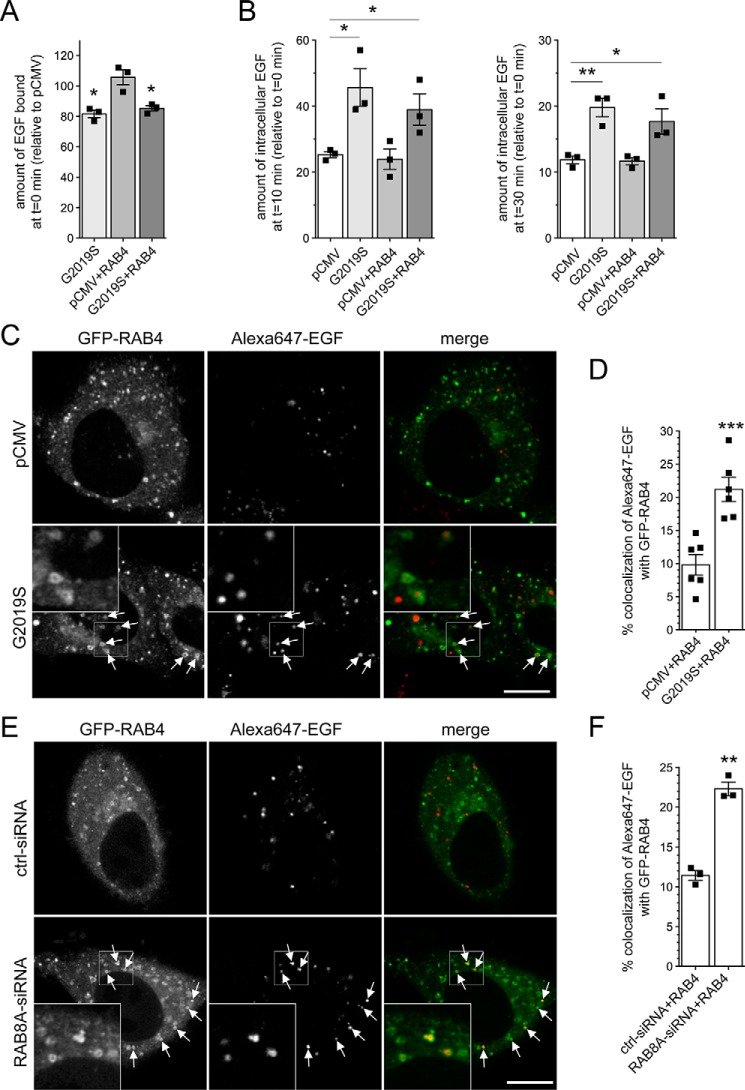
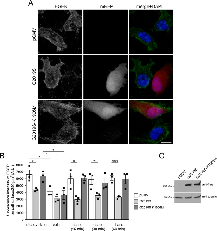
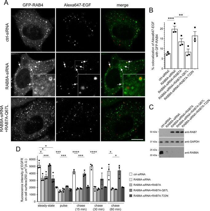
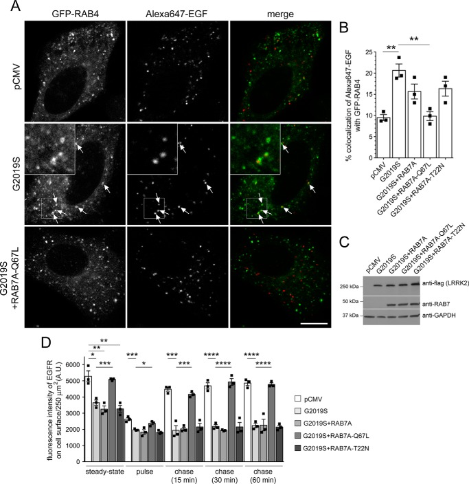
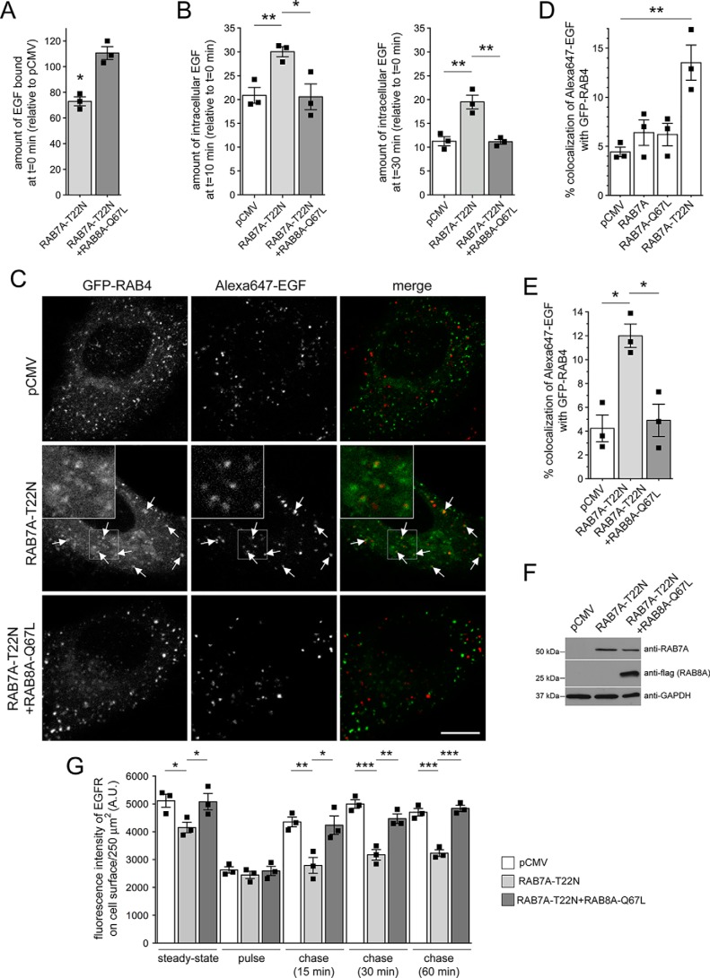
Mutations in the gene encoding for leucine-rich repeat kinase 2 (LRRK2) are a common cause of hereditary Parkinson's disease. LRRK2 regulates various intracellular vesicular trafficking pathways, including endolysosomal degradative events such as epidermal growth factor receptor (EGFR) degradation. Recent studies have revealed that a subset of RAB proteins involved in secretory and endocytic recycling are LRRK2 kinase substrates in vivo However, the effects of LRRK2-mediated phosphorylation of these substrates on membrane trafficking remain unknown. Here, using an array of immunofluorescence and pulldown assays, we report that expression of active or phosphodeficient RAB8A variants rescues the G2019S LRRK2-mediated effects on endolysosomal membrane trafficking. Similarly, up-regulation of the RAB11-Rabin8-RAB8A cascade, which activates RAB8A, also reverted these trafficking deficits. Loss of RAB8A mimicked the effects of G2019S LRRK2 on endolysosomal trafficking and decreased RAB7A activity. Expression of pathogenic G2019S LRRK2 or loss of RAB8A interfered with EGFR degradation by causing its accumulation in a RAB4-positive endocytic compartment, which was accompanied by a deficit in EGFR recycling and was rescued upon expression of active RAB7A. Dominant-negative RAB7A expression resulted in similar deficits in EGF degradation, accumulation in a RAB4 compartment, and deficits in EGFR recycling, which were all rescued upon expression of active RAB8A. Taken together, these findings suggest that, by impairing RAB8A function, pathogenic G2019S LRRK2 deregulates endolysosomal transport and endocytic recycling events.

Keywords: GTPase; Parkinson disease; RAB7A; RAB8A; Rab; early recycling compartment; endolysosome; leucine-rich repeat kinase 2 (LRRK2); lysosome; neurodegeneration; protein homeostasis; receptor endocytosis; receptor recycling.


Conflict of interest statement

The authors declare that they have no conflicts of interest with the contents of this article

Figures

Figure 1.
Figure 1.
Pathogenic G2019S LRRK2, but not a kinase-inactive G2019S-K1906M variant, causes a deficit in EGF binding and degradation. A, HeLa cells were transfected with either pCMV or cotransfected with GFP and either myc-tagged G2019S LRRK2, FLAG-tagged G2019S, or G2019S-K1906M LRRK2 as indicated; incubated with Alexa555-EGF for 30 min at 4 °C; washed to remove unbound fluorescent EGF; and fixed and processed as described under “Materials and methods.” Scale bar, 10 μm. B, quantification of surface-bound fluorescent EGF (t = 0 min) of cells transfected with the various constructs as indicated and normalized to EGF surface binding of pCMV-transfected cells (ctrl). n = 3 independent experiments. *, p < 0.05. C, HeLa cells transfected with the indicated constructs were allowed to bind Alexa555-EGF at 4 °C, washed to remove unbound fluorescent EGF, and then shifted to 37 °C for 10 min to allow for the internalization and degradation of fluorescent EGF. Scale bar, 10 μm. D, quantification of Alexa555-EGF was performed after 10 (left) and 30 min (right) upon internalization and normalized to the amount of Alexa555-EGF binding for each condition at t = 0 min, thus reflecting the percentage of internalized bound fluorescent EGF. n = 3 independent experiments. *, p < 0.05; ***, p < 0.005. E, quantification of the total number of fluorescent EGF-positive puncta per 250 μm2 upon expression of distinct constructs as indicated after 10 (left) and 30 min (right) of internalization. n = 3 independent experiments. *, p < 0.05; ***, p < 0.005. F, because the immunofluorescence signal intensity directly correlates to the size of the individual structures, signal intensity per punctum was quantified at 10 (left) and 30 min (right) upon internalization, which revealed no change among the different conditions, further indicating a deficit in EGF degradation rather than an increase in the amount of internalized fluorescent EGF per cell. G, HEK293T cells were transfected with the indicated constructs followed by analysis of endogenous EGFR expression levels. H, HEK293T cells were transfected with either pathogenic G2019S LRRK2 or with kinase-inactive G2019S-K1906M variant and serum-starved for 1 h in the presence of cycloheximide to block novel protein synthesis, and EGFR internalization was stimulated with nonlabeled EGF for the indicated time points. Cell extracts were analyzed by Western blotting for EGFR levels, and tubulin was used as a loading control. I, quantification of EGFR degradation in HEK293T cells transfected with either G2019S LRRK2 or G2019S-K1906M, at distinct time points as indicated, and with values normalized to tubulin as a loading control. n = 4 independent experiments. *, p < 0.05; **, p < 0.01; ****, p < 0.001. All error bars represent S.E.M.
Figure 2.
Figure 2.
Active RAB8A and Rabin8 rescue the LRRK2-mediated deficit in EGF binding and degradation. A, HeLa cells were transfected with either empty pCMV vector (ctrl) or the indicated RAB8A constructs followed by quantification of the amount of surface-bound fluorescent EGF. n = 4 independent experiments. *, p < 0.05. B, cells were transfected as indicated followed by quantification of internalized Alexa555-EGF in transfected cells after 10 (left) and 30 min (right) of internalization. Values are normalized to the amount of Alexa555-EGF binding at t = 0. n = 4 independent experiments. *, p < 0.05. C, cells were cotransfected with G2019S LRRK2 and the indicated RAB8A constructs, and surface-bound fluorescent EGF was quantified. n = 8 independent experiments. *, p < 0.05. D, cells were cotransfected with G2019S LRRK2 and the indicated RAB8A constructs followed by quantification of internalized Alexa555-EGF after 10 (left) and 30 min (right) of internalization. n = 8 independent experiments. ****, p < 0.001. E, cells were transfected with either empty pCMV vector (ctrl) or with Rabin8, and surface-bound fluorescent EGF was quantified. n = 3 independent experiments. F, cells were transfected as indicated followed by quantification of internalized fluorescent EGF at 10 (left) and 30 min (right). n = 3 independent experiments. G, cells were transfected with either empty pCMV vector (ctrl) or cotransfected with G2019S pathogenic LRRK2 and either pCMV vector or Rabin8 as indicated, and surface-bound fluorescent EGF was quantified. n = 3 experiments. *, p < 0.05. H, cells were transfected as indicated followed by quantification of internalized fluorescent EGF as described above. n = 3 independent experiments. ***, p < 0.005. All error bars represent S.E.M.
Figure 3.
Figure 3.
RAB11 rescues the LRRK2-mediated delay in EGFR trafficking. A, HeLa cells were cotransfected with G2019S LRRK2 and the indicated RAB11 constructs, and surface-bound fluorescent EGF was quantified. n = 3 independent experiments. *, p < 0.05. B, cells were transfected with either empty pCMV vector (ctrl) or cotransfected with G2019S LRRK2 and the indicated RAB11 constructs followed by quantification of internalized fluorescent EGF at 10 (left) and 30 min (right). n = 3 independent experiments. *, p < 0.05; **, p < 0.01; ***, p < 0.005. C, cells were transfected with either empty pCMV vector (ctrl) or the indicated RAB11 constructs, and the amount of surface-bound fluorescent EGF was quantified. n = 3 independent experiments. D, cells were transfected as indicated followed by quantification of internalized EGF at 10 (left) and 30 min (right). n = 3 independent experiments. E, cells were transfected with either empty pCMV vector (ctrl) or cotransfected with G2019S LRRK2 and the indicated RAB18 constructs, and surface-bound fluorescent EGF was quantified. n = 3 independent experiments. *, p < 0.05. F, cells were transfected as indicated followed by quantification of internalized fluorescent EGF at 10 (left) and 30 min (right). n = 3 independent experiments. ***, p < 0.005. G, same as in E, but cells were transfected with either empty pCMV vector (ctrl) or the indicated RAB18 constructs. n = 3 independent experiments. H, same as in F, but cells were transfected with either empty pCMV vector (ctrl) or the indicated RAB18 constructs. n = 3 independent experiments. All error bars represent S.E.M.
Figure 4.
Figure 4.
Phosphodeficient RAB8A, but not WT or phosphomimetic RAB8A variants, revert the LRRK2-mediated effects on EGFR trafficking. A, HeLa cells were transfected with either empty pCMV vector (ctrl) or the indicated RAB8A constructs, and surface-bound fluorescent EGF was quantified. n = 4 independent experiments. *, p < 0.05. B, cells were transfected with the indicated constructs followed by quantification of internalized fluorescent EGF. n = 4 independent experiments. *, p < 0.05. C, cells were cotransfected with G2019S LRRK2 and the indicated RAB8A constructs, and surface-bound fluorescent EGF was quantified. n = 8 independent experiments. *, p < 0.05. D, cells were transfected with the indicated constructs, and internalized fluorescent EGF was quantified at 10 (left) and 30 min (right). n = 8 independent experiments. *, p < 0.05; ***, p < 0.005; ****, p < 0.001. All error bars represent S.E.M.
Figure 5.
Figure 5.
Knockdown of RAB8A mimics the endolysosomal trafficking deficits mediated by G2019S LRRK2. A, HeLa cells were either nontransfected (−) or transfected with ctrl-siRNA or RAB8A-siRNA, and cell extracts (30 μg) were analyzed by Western blotting for RAB8A protein levels and tubulin as a loading control. B, quantification of the type of experiments depicted in A. RAB8A levels in the presence of RAB8A-siRNA were normalized to levels in the presence of ctrl-siRNA. n = 3 independent experiments. *, p < 0.05. C, cells were either left untreated (−) or transfected with ctrl-siRNA or RAB8A-siRNA, and surface-bound fluorescent EGF was quantified. n = 3 independent experiments. *, p < 0.05. D, cells were either left untreated (−) or transfected with ctrl-siRNA or RAB8A-siRNA followed by quantification of internalized fluorescent EGF at 10 (left) and 30 min (right). n = 3 independent experiments. **, p < 0.01; ****, p < 0.001. E, cells were either left untreated or cotransfected with ctrl-siRNA or RAB8A-siRNA in the absence or presence of GFP-tagged active RAB7A (RAB7A-Q67L), and surface-bound fluorescent EGF was quantified. n = 3 independent experiments. *, p < 0.05. F, cells were either left untreated or cotransfected with ctrl-siRNA or RAB8A-siRNA in the absence or presence of RAB7A-Q67L, and internalized fluorescent EGF was quantified at 10 (left) and 30 min (right). n = 3 independent experiments. *, p < 0.05; **, p < 0.01. G, cells were either treated with ctrl-siRNA or RAB8A-siRNA as indicated, and the RAB7-binding domain of RILP coupled to GST was used to pull down the GTP-bound form of RAB7 from cell lysates (300 μg). Input (10%) was run alongside pulldowns to demonstrate equal levels of total RAB7 protein in ctrl-siRNA– or RAB8A-siRNA–treated cells, and the levels of RAB8A and tubulin were analyzed on a separate gel. H, experiments of the type depicted in G were quantified, and the amount of RAB7 isolated by GST-RILP was expressed relative to input. n = 3 independent experiments. ***, p < 0.005. I, cells were either treated with ctrl-siRNA or RAB8A-siRNA as indicated, and a conformation-specific antibody was used to immunoprecipitate active RAB7 from cell lysates (2 mg). As a positive control, ctrl-siRNA–treated cell extracts were incubated with 100 μm GTPγS to activate RAB7A before immunoprecipitation. Input (1%) was run alongside pulldowns to demonstrate equal levels of total RAB7 protein in ctrl-siRNA– or RAB8A-siRNA–treated cells, and the levels of RAB8A and tubulin were analyzed on a separate gel. All error bars represent S.E.M.
Figure 6.
Figure 6.
Pathogenic LRRK2 or knockdown of RAB8A causes accumulation of EGF in a RAB4-positive endocytic compartment. A, HeLa cells were transfected with either empty pCMV vector or pathogenic LRRK2 or cotransfected with GFP-tagged RAB4, and surface-bound fluorescent EGF was quantified. n = 3 independent experiments. *, p < 0.05. B, cells were transfected as indicated followed by quantification of internalized fluorescent EGF at 10 (left) and 30 min (right). n = 3 independent experiments. *, p < 0.05; **, p < 0.01. C, example of HeLa cells cotransfected with GFP-RAB4 and either empty pCMV vector or pathogenic LRRK2. Live pictures were taken 20 min upon fluorescent EGF internalization, and arrows point to GFP-RAB4–positive vesicles containing Alexa647-EGF. Scale bar, 10 μm. D, quantification of colocalization of Alexa647-EGF with GFP-RAB4 (Manders' coefficient 1 × 100) from 15–20 cells per experiment. n = 6 independent experiments. ***, p < 0.005. E, example of HeLa cells cotransfected with GFP-RAB4 and either ctrl-siRNA or RAB8A-siRNA. Live pictures were taken as described above. Arrows point to GFP-RAB4–positive vesicles containing Alexa647-EGF. Scale bar, 10 μm. F, quantification of colocalization of Alexa647-EGF with GFP-RAB4 (Manders' coefficient 1 × 100) from 15–20 cells per experiment. n = 3 independent experiments. **, p < 0.01. All error bars represent S.E.M.
Figure 7.
Figure 7.
Pathogenic G2019S LRRK2 causes a deficit in EGFR recycling. A, example of HeLa cells transfected with pCMV or cotransfected with mRFP and either G2019S or kinase-inactive G2019S-K1906M LRRK2 and stained with an antibody against the extracellular domain of the EGFR in the absence of permeabilization to visualize only surface EGFR. Scale bar, 10 μm. B, quantification of fluorescence intensity of surface levels of EGFR at t = 0 min (steady-state), upon triggering internalization of the EGFR (pulse), or upon chase for various time points to assess recycling rates (chase) as described under “Materials and methods.” n = 3 independent experiments. *, p < 0.05; ***, p < 0.005. C, HeLa cells were transfected as indicated, and cell extracts (30 μg) were analyzed by Western blotting for FLAG-tagged LRRK2 levels and tubulin as a loading control. A.U., arbitrary units. All error bars represent S.E.M.
Figure 8.
Figure 8.
Accumulation of EGF in a RAB4-positive endocytic compartment and deficits in EGFR recycling due to knockdown of RAB8A are rescued by active RAB7A expression. A, example of HeLa cells cotransfected with GFP-RAB4 and either ctrl-siRNA or RAB8A-siRNA with or without RAB7A-Q67L expression as indicated. Live pictures were taken 20 min upon fluorescent EGF internalization, and arrows point to GFP-RAB4–positive vesicles containing Alexa647-EGF. An independent picture (543 HeNe laser line) was acquired to confirm coexpression of the distinct mRFP-tagged RAB7A constructs in all cases. Scale bar, 10 μm. B, quantification of colocalization of Alexa647-EGF with GFP-RAB4 and either ctrl-siRNA or RAB8A-siRNA in the presence or absence of distinct RAB7A constructs as indicated (Manders' coefficient 1 × 100) from 15–20 cells per experiment. n = 3 independent experiments. **, p < 0.01; ***, p < 0.005. C, HeLa cells were treated with ctrl-siRNA or RAB8A-siRNA as indicated and transfected with the indicated RAB7A constructs, and cell extracts (30 μg) were analyzed by Western blotting for RAB8A protein levels, mRFP-RAB7A protein levels (anti-RAB7 antibody), and GAPDH as a loading control. D, HeLa cells were treated with either ctrl-siRNA or RAB8A-siRNA as indicated with or without cotransfection with the indicated RAB7A constructs. EGFR recycling assays were performed as described under “Materials and methods,” revealing a deficit in EGFR surface levels and EGFR recycling upon RAB8A-siRNA, which was rescued upon expression of active RAB7A. n = 3 independent experiments. *, p < 0.05; ***, p < 0.005; ****, p < 0.001. A.U., arbitrary units. All error bars represent S.E.M.
Figure 9.
Figure 9.
Accumulation of EGF in a RAB4-positive endocytic compartment and deficits in EGFR recycling due to G2019S LRRK2 expression are rescued by active RAB7A expression. A, example of HeLa cells cotransfected with GFP-RAB4 and either empty pCMV vector or pathogenic LRRK2 with or without RAB7A-Q67L expression as indicated. Live pictures were taken 20 min upon fluorescent EGF internalization, and arrows point to GFP-RAB4–positive vesicles containing Alexa647-EGF. An independent picture (543 HeNe laser line) was acquired to confirm coexpression of the distinct mRFP-tagged RAB7A constructs in all cases. Scale bar, 10 μm. B, quantification of colocalization of Alexa647-EGF with GFP-RAB4 in cells coexpressing empty pCMV vector or G2019S LRRK2 in the presence or absence of mRFP-tagged RAB7A constructs as indicated (Manders' coefficient 1 × 100) from 15–20 cells per experiment. n = 3 independent experiments. **, p < 0.01. C, HeLa cells were transfected with the indicated constructs, and cell extracts (30 μg) were analyzed by Western blotting for FLAG-tagged G2019S-LRRK2, mRFP-RAB7A protein levels (anti-RAB7 antibody), and GAPDH as a loading control. D, HeLa cells were transfected with either empty pCMV vector or pathogenic G2019S LRRK2 in the presence or absence of mRFP-tagged RAB7A constructs as indicated, and EGFR surface levels and EGFR recycling were determined at the indicated time points. n = 3 independent experiments. *, p < 0.05; **, p < 0.01; ***, p < 0.005; ****, p < 0.001. A.U., arbitrary units. All error bars represent S.E.M.
Figure 10.
Figure 10.
Expression of dominant-negative RAB7A causes defects in EGFR trafficking, accumulation of EGF in a RAB4-positive endocytic compartment, and deficits in EGFR recycling, which are reversed upon active RAB8A expression. A, HeLa cells were transfected with either empty pCMV vector (ctrl) or with dominant-negative RAB7A (RAB7A-T22N) in the presence or absence of active RAB8A (RAB8A-Q67L), and surface-bound fluorescent EGF was quantified. n = 3 independent experiments. *, p < 0.05. B, cells were transfected with the indicated constructs followed by quantification of internalized fluorescent EGF at 10 (left) and 30 min (right). n = 3 independent experiments. *, p < 0.05; **, p < 0.01. C, example of HeLa cells cotransfected with GFP-RAB4 and either mRFP-RAB7A-T22N or mRFP-RAB7A-T22N and FLAG-tagged RAB8A-Q67L as indicated. Live pictures were taken 20 min upon fluorescent EGF internalization, and arrows point to GFP-RAB4–positive vesicles containing Alexa647-EGF. An independent picture (543 HeNe laser line) was acquired to confirm coexpression of the mRFP-tagged RAB7A constructs in all cases. Scale bar, 10 μm. D, quantification of colocalization of Alexa647-EGF with GFP-RAB4 in the presence or absence of the distinct RAB7A constructs as indicated (Manders' coefficient 1 × 100) from 15–20 cells per experiment. n = 3 independent experiments. **, p < 0.01. E, quantification of colocalization of Alexa647-EGF with GFP-RAB4 in the presence or absence of RAB7A-T22N and RAB8A-Q67L constructs as indicated (Manders' coefficient 1 × 100) from 15–20 cells per experiment. n = 3 independent experiments. *, p < 0.05. F, HeLa cells were transfected with the indicated constructs, and cell extracts (30 μg) were analyzed by Western blotting for mRFP-tagged RAB7A-T22N, FLAG-tagged RAB8A-Q67L, and GAPDH as a loading control. G, HeLa cells were transfected with either empty pCMV vector or dominant-negative RAB7A-T22N in the absence or presence of RAB8A-Q67L, and EGFR surface levels and EGFR recycling were determined at the indicated time points. n = 3 independent experiments. *, p < 0.05; **, p < 0.01; ***, p < 0.005. A.U., arbitrary units. All error bars represent S.E.M.

Similar articles

Cited by

References

    1. Paisán-Ruíz C., Jain S., Evans E. W., Gilks W. P., Simón J., van der Brug M., López de Munain A., Aparicio S., Gil A. M., Khan N., Johnson J., Martinez J. R., Nicholl D., Carrera I. M., Pena A. S., et al. (2004) Cloning of the gene containing mutations that cause PARK8-linked Parkinson's disease. Neuron 44, 595–600 10.1016/j.neuron.2004.10.023 - DOI - PubMed
    1. Zimprich A., Biskup S., Leitner P., Lichtner P., Farrer M., Lincoln S., Kachergus J., Hulihan M., Uitti R. J., Calne D. B., Stoessl A. J., Pfeiffer R. F., Patenge N., Carbajal I. C., Vieregge P., et al. (2004) Mutations in LRRK2 cause autosomal-dominant parkinsonism with pleomorphic pathology. Neuron 44, 601–607 10.1016/j.neuron.2004.11.005 - DOI - PubMed
    1. Healy D. G., Falchi M., O'Sullivan S. S., Bonifati V., Durr A., Bressman S., Brice A., Aasly J., Zabetian C. P., Goldwurm S., Ferreira J. J., Tolosa E., Kay D. M., Klein C., Williams D. R., et al. (2008) Phenotype, genotype, and worldwide genetic penetrance of LRRK2-associated Parkinson's disease: a case-control study. Lancet Neurol 7, 583–590 10.1016/S1474-4422(08)70117-0 - DOI - PMC - PubMed
    1. Cookson M. R., and Bandmann O. (2010) Parkinson's disease: insights from pathways. Hum. Mol. Genet. 19, R21–R27 10.1093/hmg/ddq167 - DOI - PMC - PubMed
    1. Greggio E., Jain S., Kingsbury A., Bandopadhyay R., Lewis P., Kaganovich A., van der Brug M. P., Beilina A., Blackinton J., Thomas K. J., Ahmad R., Miller D. W., Kesavapany S., Singleton A., Lees A., et al. (2006) Kinase activity is required for the toxic effects of mutant LRRK2/dardarin. Neurobiol. Dis. 23, 329–341 10.1016/j.nbd.2006.04.001 - DOI - PubMed

Publication types

MeSH terms