Review
. 2019 Feb 1;294(5):1643-1651.
doi: 10.1074/jbc.TM118.004156.

Proteases: History, discovery, and roles in health and disease

Affiliations
Review

Proteases: History, discovery, and roles in health and disease

Judith S Bond. J Biol Chem. .

Abstract

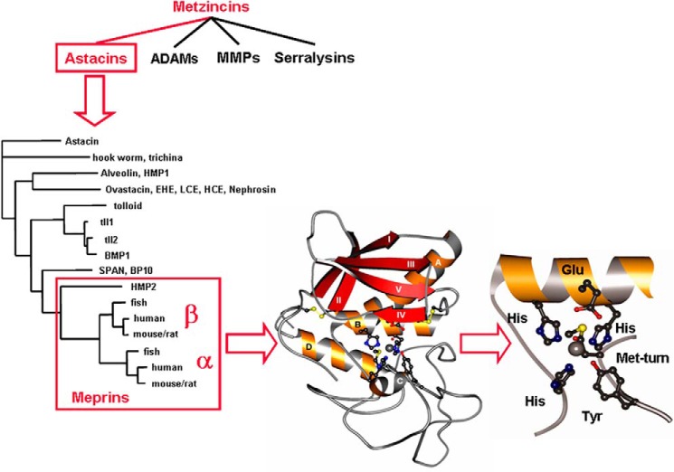
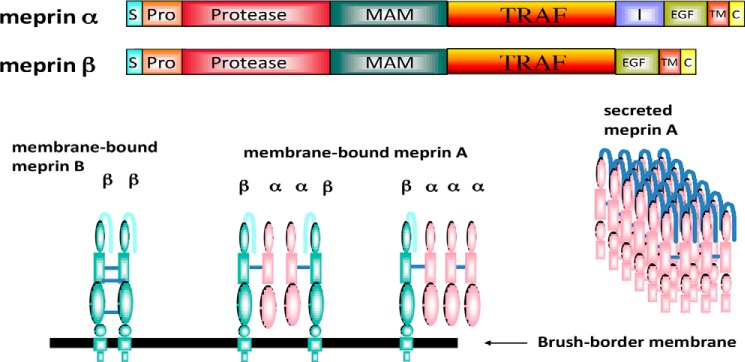
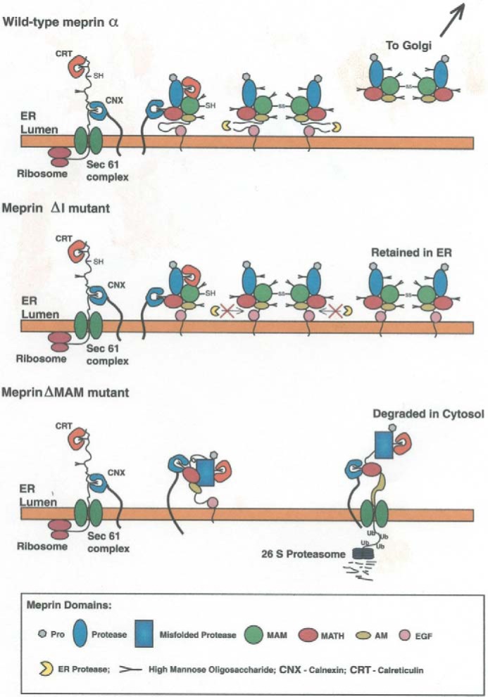
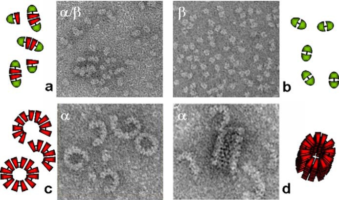
The Journal of Biological Chemistry (JBC) has been a major vehicle for disseminating and recording the discovery and characterization of proteolytic enzymes. The pace of discovery in the protease field accelerated during the 1971-2010 period that Dr. Herb Tabor served as the JBC's editor-in-chief. When he began his tenure, the fine structure and kinetics of only a few proteases were known; now thousands of proteases have been characterized, and over 600 genes for proteases have been identified in the human genome. In this review, besides reflecting on Dr. Tabor's invaluable contributions to the JBC and the American Society for Biochemistry and Molecular Biology (ASBMB), I endeavor to provide an overview of the extensive history of protease research, highlighting a few discoveries and roles of proteases in vivo In addition, metalloproteinases, particularly meprins of the astacin family, will be discussed with regard to structural characteristics, regulation, mechanisms of action, and roles in health and disease. Proteases and protein degradation play crucial roles in living systems, and I briefly address future directions in this highly diverse and thriving research area.

Keywords: astacins; meprins; metalloprotease; metalloproteinase; protein complex; protein degradation; protein domain; proteinase.


Conflict of interest statement

The author declares that she has no conflicts of interest with the contents of this article

Figures

Figure 1.
Figure 1.
Metzinicins, astacins, and meprins. The metzincin superfamily includes the astacins, the ADAMs, the MMPs, the serralysins, and the papalysins and leishmanolysins (last two families not shown). Over 180 individual astacins have been identified in animals and bacteria, and several examples are shown, including the crayfish astacin, fly and human tolloids, and the meprins from hydra (HMP2), zebrafish, human, and mouse/rat. The ribbon diagram shows that the protease domain of meprins consists of five-stranded β-sheets, three α-helices, and a coil structure in the lower subdomain. The zinc (gray sphere) is pentacoordinated by three histidines of the motif HEXXHXXGXXH, a water molecule, and a tyrosine positioned by the Met-turn. Adapted from Ref. . This research was originally published in Molecular Aspects of Medicine. Sterchi, E. E., Stöcker, W., and Bond, J. S. Meprins, membrane-bound and secreted astacin metalloproteinases. Molecular Aspects of Medicine. 2008; 29, 309–328. © Elsevier Ltd.
Figure 2.
Figure 2.
Domain and oligomeric structure of meprins α and β. Domains are as follows: S (signal sequence), Pro (prosequence), protease, catalytic domain, MAM (meprin), A5 protein, protein-tyrosine phosphatase μ, TRAF homology, I (inserted), EGF (epidermal growth factor-like), TM (transmembrane-spanning), and C (cytoplasmic). During maturation, the meprin α subunit is cleaved in the I domain, separating the subunit from the membrane. As a result, three isoforms of meprin exist: membrane-bound meprin B (a homodimer of β subunits), membrane-bound meprin A (heterotetramers of α and β subunits, found in ratios of α2β2 and α1β3), and secreted meprin A (homomeric multimers of α subunit dimers). The secreted forms of meprin α dimers tend to self-associate and form large multimers (1–6 MDa) extracellularly.
Figure 3.
Figure 3.
Intracellular trafficking of WT meprin α subunits and mutants. WT meprin α is secreted from cells after maturation in the endoplasmic reticulum (ER) and Golgi; if the I domain is deleted by site-directed mutagenesis (ΔI mutant), the subunit is retained in the endoplasmic reticulum/cis-Golgi; if the MAM domain is deleted (ΔMAM mutant) the subunit misfolds, and this triggers retrograde transport to the cytosol and degradation by the proteasome (64).
Figure 4.
Figure 4.
Oligomerization of meprin A and B. Shown are electron micrographs of rat meprin A and B expressed in human embryonic kidney 293 cells. Negatively stained samples of various isoforms are shown. Shown clockwise from left to right are the following: heteromeric meprin A containing tetramers of α and β subunits (a); homomeric meprin B (dimers of β subunits) (b); latent homomeric meprin A containing homodimers of meprin α subunits that associate noncovalently to form crescents, tubes, and spirals containing up to 100 subunits (d); and activated homomeric meprin A forming primarily rings and crescents containing about 10–12 subunits (c). Adapted from Refs. and . This research was originally published in Molecular Aspects of Medicine. Sterchi, E. E., Stöcker, W., and Bond, J. S. Meprins, membrane-bound and secreted astacin metalloproteinases. Molecular Aspects of Medicine. 2008; 29, 309–328. © Elsevier Ltd. and the Journal of Biological Chemistry. Bertenshaw, G. P., Norcum, M. T., and Bond, J. S. Structure of homo- and hetero-oligomeric meprin metalloproteases: dimers, tetramers, and high molecular mass multimers. Journal of Biological Chemistry. 2003; 278, 2522–2532. © the American Society for Biochemistry and Molecular Biology.

Similar articles

Cited by

References

    1. Levene P. A. (1905) The cleavage products of proteoses. J. Biol. Chem. 1, 45–58
    1. Northrup J. H. (1930) Crystalline pepsin. I. Isolation and tests of purity. J. Gen. Physiol. 13, 739–766 10.1085/jgp.13.6.739 - DOI - PMC - PubMed
    1. Robertson T. B. (1907) Note on the synthesis of a protein through the action of pepsin. J. Biol. Chem. 3, 95–99
    1. Northrop J. H., and Kunitz M. (1931) Isolation of protein crystals processing tryptic activity. Science 73, 262–263 10.1126/science.73.1888.262 - DOI - PubMed
    1. Robertson T. B. (1907) Studies in the chemistry of the ion-proteid compounds: IV. On some chemical properties of casein and their possible relation to the chemical behavior of other protein bodies, with especial reference to hydrolysis of casein by trypsin. J. Biol. Chem. 2, 317–383

Substances

LinkOut - more resources