

Medial canthoplasty for the management of exposure keratopathy
- PMID: 30710111
- PMCID: PMC6707211
- DOI: 10.1038/s41433-019-0347-9
Medial canthoplasty for the management of exposure keratopathy
Abstract
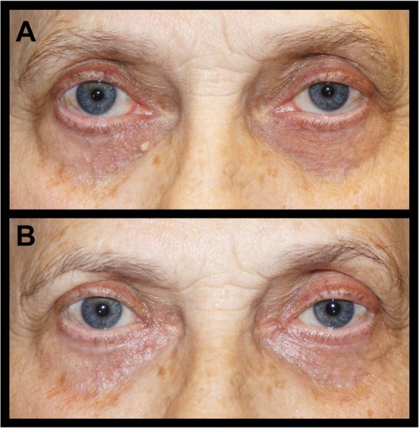
Purpose: To report the surgical technique and functional outcomes of the medial canthoplasty for the treatment of exposure keratopathy.
Patients/methods: An IRB approved, retrospective review of patients who underwent medial canthoplasty for exposure keratopathy was performed. Patient demographics, reported symptoms, and clinical examination findings were collected and analyzed from pre-operative and follow-up visits.
Results: The study included 73 consecutive cases in which the medial canthoplasty was performed in patients with exposure keratopathy. The average follow-up period was 7.9 months (median: 4.7 months; range: 1-150 months). Complete or partial improvement in ocular symptoms (dryness; pain/irritation; tearing) was achieved in 95% (69/73). Clinically, 85% (41/48) of patients demonstrated a post-operative reduction in lagophthalmos and 90% (60/67) showed improvement in ocular surface findings. Complications were rare (1/73) and reversal of medial canthoplasty was not required in any case.
Conclusions: The medial canthoplasty appears to be a safe and effective technique to narrow the palpebral fissure, provide lower eyelid support, and improve keratopathy. It is an uncomplicated procedure that may be considered for the treatment of exposure keratopathy caused by facial paralysis and lower eyelid malposition.
Conflict of interest statement
The authors declare that they have no conflict of interest.
Figures

Similar articles
-
Palpebral spring in the management of lagophthalmos and exposure keratopathy secondary to facial nerve palsy.Ophthalmic Plast Reconstr Surg. 2009 Jul-Aug;25(4):270-5. doi: 10.1097/IOP.0b013e3181ab6f08. Ophthalmic Plast Reconstr Surg. 2009. PMID: 19617783
-
Technique and Results of Permanent Medial Tarsorrhaphy for Complex Eyelid Malposition.Ophthalmic Plast Reconstr Surg. 2019 Mar/Apr;35(2):197-201. doi: 10.1097/IOP.0000000000001282. Ophthalmic Plast Reconstr Surg. 2019. PMID: 30856627
-
Re: "Palpebral spring in the management of lagophthalmos and exposure keratopathy secondary to facial nerve palsy".Ophthalmic Plast Reconstr Surg. 2010 Nov-Dec;26(6):499-500. doi: 10.1097/IOP.0b013e3181cc872e. Ophthalmic Plast Reconstr Surg. 2010. PMID: 20639787 No abstract available.
-
[Conceptual basics of paralytic lagophthalmos correction].Vestn Oftalmol. 2013 Sep-Oct;129(5):92-6. Vestn Oftalmol. 2013. PMID: 24261285 Review. Russian.
-
Lateral canthoplasty.Ophthalmic Plast Reconstr Surg. 2003 Sep;19(5):345-52. doi: 10.1097/01.IOP.0000087069.83107.A4. Ophthalmic Plast Reconstr Surg. 2003. PMID: 14506418 Review. No abstract available.
Cited by
-
A Review of Clinical Outcomes, Owner Understanding and Satisfaction following Medial Canthoplasty in Brachycephalic Dogs in a UK Referral Setting (2016-2021).Animals (Basel). 2023 Jun 19;13(12):2032. doi: 10.3390/ani13122032. Animals (Basel). 2023. PMID: 37370542 Free PMC article.
-
Ophthalmological Care of Patients With Craniofacial Disorders.J Pediatr Neurosci. 2022 Sep;17(Suppl 1):S61-S66. doi: 10.4103/jpn.JPN_45_22. Epub 2022 Sep 19. J Pediatr Neurosci. 2022. PMID: 36388012 Free PMC article. Review.
References
-
- Piskiniene R. Eyelid malposition: lower lid entropion and ectropion. Medicine (Kaunas) 2006;42:881–4. - PubMed
MeSH terms
LinkOut - more resources
Full Text Sources
Medical
