

Structural and biomechanical changes to dentin extracellular matrix following chemical removal of proteoglycans
- PMID: 30710179
- PMCID: PMC6559829
- DOI: 10.1007/s10266-018-00408-0
Structural and biomechanical changes to dentin extracellular matrix following chemical removal of proteoglycans
Abstract
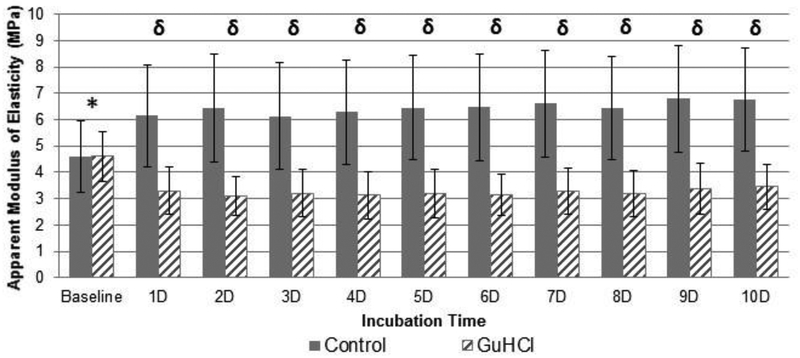
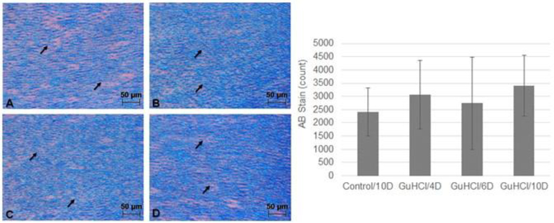
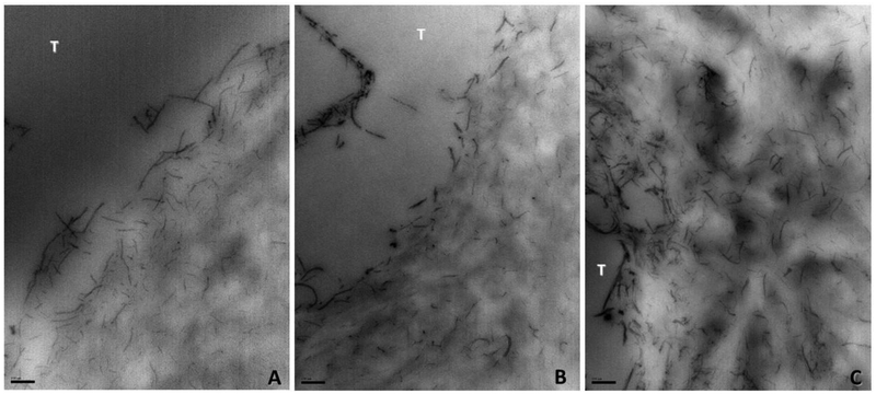
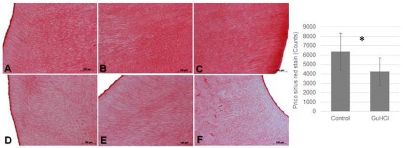
Proteoglycans are biomacromolecules with significant biomineralization and structural roles in the dentin extracellular matrix. This study comprehensively assessed the mechanical properties and morphology of the dentin extracellular matrix following chemical removal of proteoglycans to elucidate the structural roles of proteoglycans in dentin. Dentin extracellular matrix was prepared from extracted teeth after complete tissue demineralization. Chemical removal of proteoglycans was carried-out using guanidine hydrochloride for up to 10 days. The removal of proteoglycans was determined by dimethylmethylene blue colorimetric assay and histological staining analyses using transmission electron microscopy and optical microscopy. The modulus of elasticity of dentin matrix was determined by a 3-point bending test method. Partial removal of proteoglycans induced significant modifications to the dentin matrix, particularly to type I collagen. Removal of proteoglycans significantly decreased the modulus of elasticity of dentin extracellular matrix (p < 0.0001). In conclusion, the subtle disruption of proteoglycans induces pronounced changes to the collagen network packing and the bulk modulus of elasticity of dentin matrix.
Keywords: Collagen; Dentin; Histology; Modulus of elasticity; Proteoglycans.
Conflict of interest statement
Conflict of Interest
The authors state no conflict of interest with the content of the manuscript.
Figures
Similar articles
-
Role of proteoglycans on the biochemical and biomechanical properties of dentin organic matrix.Arch Oral Biol. 2017 Oct;82:203-208. doi: 10.1016/j.archoralbio.2017.06.020. Epub 2017 Jun 16. Arch Oral Biol. 2017. PMID: 28651092 Free PMC article.
-
The stoic tooth root: how the mineral and extracellular matrix counterbalance to keep aged dentin stable.Acta Biomater. 2022 Jan 15;138:351-360. doi: 10.1016/j.actbio.2021.10.051. Epub 2021 Nov 2. Acta Biomater. 2022. PMID: 34740855 Free PMC article.
-
Insights into the structure and composition of the peritubular dentin organic matrix and the lamina limitans.Micron. 2012 Feb;43(2-3):229-36. doi: 10.1016/j.micron.2011.08.003. Epub 2011 Aug 12. Micron. 2012. PMID: 21890367
-
The dentin substrate: structure and properties related to bonding.J Dent. 1997 Nov;25(6):441-58. doi: 10.1016/s0300-5712(96)00065-6. J Dent. 1997. PMID: 9604576 Review.
-
[Proteoglycans-phospholipids interactions: roles in dentine mineralization].C R Seances Soc Biol Fil. 1993;187(2):210-22. C R Seances Soc Biol Fil. 1993. PMID: 8019902 Review. French.
Cited by
-
Stability and remineralization of proteoglycan-infused dentin substrate.Dent Mater. 2021 Nov;37(11):1724-1733. doi: 10.1016/j.dental.2021.09.003. Epub 2021 Sep 15. Dent Mater. 2021. PMID: 34538503 Free PMC article.
-
On the bulk biomechanical behavior of densely cross-linked dentin matrix: The role of induced-glycation, regional dentin sites and chemical inhibitor.J Mech Behav Biomed Mater. 2020 Mar;103:103589. doi: 10.1016/j.jmbbm.2019.103589. Epub 2019 Dec 9. J Mech Behav Biomed Mater. 2020. PMID: 32090918 Free PMC article.
-
From Translation to Protein Degradation as Mechanisms for Regulating Biological Functions: A Review on the SLRP Family in Skeletal Tissues.Biomolecules. 2020 Jan 3;10(1):80. doi: 10.3390/biom10010080. Biomolecules. 2020. PMID: 31947880 Free PMC article. Review.
References
-
- Bertassoni LE, Swain MV. The contribution of proteoglycans to the mechanical behavior of mineralized tissues. J Mech Behav Biomed Mater 2014;38:91–104. - PubMed
-
- Imbeni V, Kruzicm JJ, Marshall GW, Marshall SJ, Ritchie RO. The dentin-enamel junction and the fracture of human teeth. Nat Mater 2005;4:229–32. - PubMed
-
- Dechichi P, Biffi JC, Moura CC, de Ameida AW. A model of the early mineralization process of mantle dentin. Micron 2007;38:486–91. - PubMed
MeSH terms
Substances
Grants and funding
LinkOut - more resources
Full Text Sources
