Review
. 2019 Jul;24(4):575-586.
doi: 10.1007/s10741-019-09770-9.

The relevance of microRNA in post-infarction left ventricular remodelling and heart failure

Affiliations
Review

The relevance of microRNA in post-infarction left ventricular remodelling and heart failure

Mieczysław Dutka et al. Heart Fail Rev. 2019 Jul.

Abstract

Myocardial infarction and post-infarction left ventricular remodelling involve a high risk of morbidity and mortality. For this reason, ongoing research is being conducted in order to learn the mechanisms of unfavourable left ventricular remodelling following a myocardial infarction. New biomarkers are also being sought that would allow for early identification of patients with a high risk of post-infarction remodelling and dysfunction of the left ventricle. In recent years, there has been ever more experimental data that confirms the significance of microRNA in cardiovascular diseases. It has been confirmed that microRNAs are stable in systemic circulation, and can be directly measured in patients' blood. It has been found that significant changes occur in the concentrations of various types of microRNA in myocardial infarction and heart failure patients. Various types of microRNA are also currently being intensively researched in terms of their usefulness as markers of cardiomyocyte necrosis, and predictors of the post-infarction heart failure development. This paper is a summary of the current knowledge on the significance of microRNA in post-infarction left ventricular remodelling and heart failure.

Keywords: Heart failure; Left ventricular remodelling; MicroRNA; Myocardial fibrosis; Myocardial infarction.


Conflict of interest statement

The authors declare that they have no conflict of interest.

Figures
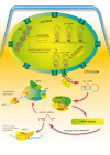
Fig. 1
Fig. 1
MicroRNA biogenesis. The first stage of biogenesis of microRNA is the transcription of miRNA genes performed in the nucleus with the participation of RNA polymerase II. This process results in the creation of pri-miRNAs. At the next stage, the pri-miRNAs are cleaved into precursor miRNAs (pre-miRNAs). This happens with the participation of the Drosha enzyme in association with the DGCR8 protein (DiGeorge syndrome Critical Region 8). This complex creates pre-miRNA. There is also an alternative pathway by the direct splicing of mRNA introns, bypassing the Drosha processing. The pre-miRNA molecules formed in this way are called mirtrons. Both these types of molecules (pre-miRNAs and mirtrons) are actively transported to the cytoplasm by Exportin-5. In the cytoplasm, the next stage of the maturation of pre-miRNA takes place. At this stage, the Dicer enzyme together with TRBP2 (transactivating response RNA-binding protein) creates the double-stranded miRNA duplexes. Each of these duplexes has a functional miRNA leading strand and a passenger strand. Next, the duplex is unwound, and the passenger strand is degraded. From this moment, the leading strand, as a mature miRNA, can enter the miRISC (miRNA-induced silencing complex) by connecting with Argonaute proteins. Other abbreviations: PABP poly A binding protein, CCR4 C-C chemokine receptor type 4, AGO-1 Argonaute protein 1, AGO-2 Argonaute protein 2
Fig. 2
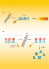
Fig. 2
The mechanisms of microRNA action. a In normal conditions, without any microRNAs, protein synthesis functions correctly. b When microRNAs are present the protein synthesis is blocked by one of two ways. The first process (rarely seen in humans) needs perfect complementarity between the microRNA and the mRNA. When the complex microRNA–mRNA is created, the degradation of mRNA starts, which blocks protein synthesis. The second process (often seen in animal cells) does not need perfect complementarity between the microRNA and the mRNA. In this process, the microRNA binds to mRNA in the 3′UTR and inhibits translation, which blocks protein synthesis

References

    1. Ueland T, Yndestad A, Dahl CP, Gullestad L, Aukrust P. TNF revisited: osteoprotegerin and TNF – related molecules in heart failure. Curr Heart Fail Rep. 2012;9:92–100. doi: 10.1007/s11897-012-0088-6. - DOI - PubMed
    1. Secchiero P, Corallini F, Beltrami A, Ceconi C, Bonasia V, et al. An imbalanced OPG/TRAIL ratio is associated to severe acute myocardial infarction. Atherosclerosis. 2010;210:274–277. doi: 10.1016/j.atherosclerosis.2009.11.005. - DOI - PubMed
    1. Bjerre M, Munk K, Sloth AD, Nielsen ST, Flyvbjerg A, et al. High osteoprotegerin levels predict MACCE in STEMI patients, but are not assotiated with myocardial salvage. Scand Cardiovasc J Vol. 2014;48:209–215. doi: 10.3109/14017431.2014.917767. - DOI - PubMed
    1. Pedersen S, Mogelvang R, Bjerre M, Frystyk J, Flyvbjerg A, Galatius S, Sørensen TB, Iversen A, Hvelplund A, Jensen JS. Osteoprotegerin predicts long-term outcome in patients with ST-segment elevation myocardial infarction treated with primary percutaneous coronary intervention. Cardiology. 2012;123:31–38. doi: 10.1159/000339880. - DOI - PubMed
    1. Corallini F, Secchiero P, Beltrami AP, CesselliD PE, et al. TNF –alfa modulates the migratory response of mesenchymal stem cells to TRAIL. Cell Mol Life Sci. 2012;67:1307–1314. doi: 10.1007/s00018-009-0246-5. - DOI - PMC - PubMed