

S100 Proteins in the Innate Immune Response to Pathogens
- PMID: 30710280
- PMCID: PMC6475579
- DOI: 10.1007/978-1-4939-9030-6_18
S100 Proteins in the Innate Immune Response to Pathogens
Abstract
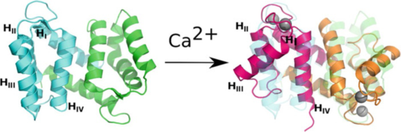
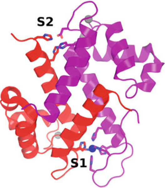
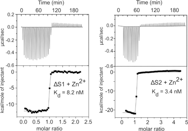
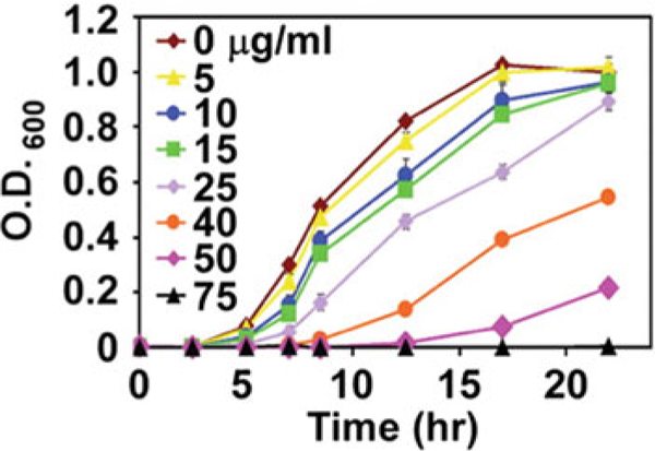
S100 proteins are distinct dimeric EF-hand Ca2+-binding proteins that can bind Zn2+, Mn2+, and other transition metals with high affinity at two sites in the dimer interface. Certain S100 proteins, including S100A7, S100A12, S100A8, and S100A9, play key roles in the innate immune response to pathogens. These proteins function via a "nutritional immunity" mechanism by depleting essential transition metals in the infection that are required for the invading organism to grow and thrive. They also act as damage-associated molecular pattern ligands, which activate pattern recognition receptors (e.g., Toll-like receptor 4, RAGE) that mediate inflammation. Here we present protocols for these S100 proteins for high-level production of recombinant protein, measurement of binding affinities using isothermal titration calorimetry, and an assay of antimicrobial activity.
Keywords: Antimicrobial growth assay; Calprotectin; Host-pathogen interaction; Inflammatory response; Isothermal titration calorimetry; Metal binding; Nutritional immunity; Protein expression; Protein purification; S100 proteins; S100A12; S100A7; S100A8; S100A9.
Figures
References
-
- Nelson MR, Chazin WJ (1998) Structures of EF-hand Ca(2+)-binding proteins: diversity in the organization, packing and response to Ca2+ binding. Biometals 11(4):297–318 - PubMed
-
- Donato R (2003) Intracellular and extracellular roles of S100 proteins. Microsc Res Tech 15:540–551 - PubMed
-
- Hunter MJ, Chazin WJ (1998) High level expression and dimer characterization of the S100 EF-hand proteins, migration inhibitory factor-related proteins 8 and 14. J Biol Chem 273:12427–12435 - PubMed
Publication types
MeSH terms
Substances
Grants and funding
LinkOut - more resources
Full Text Sources
Research Materials
Miscellaneous
