. 2019:22:101688.
doi: 10.1016/j.nicl.2019.101688. Epub 2019 Jan 25.

Anatomical features of primary brain tumors affect seizure risk and semiology

Affiliations

Anatomical features of primary brain tumors affect seizure risk and semiology

Kevin Akeret et al. Neuroimage Clin. 2019.

Abstract

Objective: An epileptic seizure is the most common clinical manifestation of a primary brain tumor. Due to modern neuroimaging, detailed anatomical information on a brain tumor is available early in the diagnostic process and therefore carries considerable potential in clinical decision making. The goal of this study was to gain a better understanding of the relevance of anatomical tumor characteristics on seizure prevalence and semiology.

Methods: We reviewed prospectively collected clinical and imaging data of all patients operated on a supratentorial intraparenchymal primary brain tumor at our department between January 2009 and December 2016. The effect of tumor histology, anatomical location and white matter infiltration on seizure prevalence and semiology were assessed using uni- and multivariate analyses.

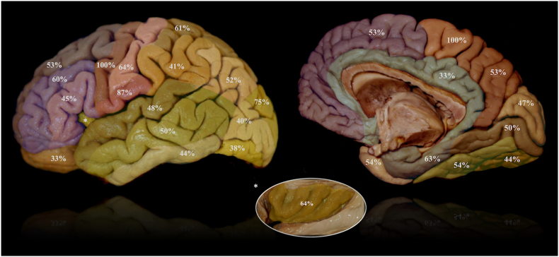
Results: Of 678 included patients, 311 (45.9%) presented with epileptic seizures. Tumor location within the central lobe was associated with higher seizure prevalence (OR 4.67, 95% CI: 1.90-13.3, p = .002), especially within the precentral gyrus or paracentral lobule (100%). Bilateral extension, location within subcortical structures and invasion of deeper white matter sectors were associated with a lower risk (OR 0.45, 95% CI: 0.25-0.78; OR 0.10, 95% CI: 0.04-0.21 and OR 0.39, 95% CI: 0.14-0.96, respectively). Multivariate analysis revealed the impact of a location within the central lobe on seizure risk to be highly significant and more relevant than histopathology (OR: 4.79, 95% CI: 1.82-14.52, p = .003). Seizures due to tumors within the central lobe differed from those of other locations by lower risk of secondary generalization (p < .001).

Conclusions: Topographical lobar and gyral location, as well as extent of white matter infiltration impact seizure risk and semiology. This finding may have a high therapeutic potential, for example regarding the use of prophylactic antiepileptic therapy.

Keywords: Anatomy; Brain tumor; Central lobe; Epilepsy; Glioma; Histology; Seizures; Topography; White matter sectors.


Figures

Unlabelled Image
Graphical abstract
Fig. 1
Fig. 1
White matter sectors. The left figure schematically shows the white matter classification used (Yasargil, 1994). It is based on purely anatomical-morphological criteria and follows a dichotomic centrifugal principle: the lobar white matter sector (IV) divides into gyral sectors (III), followed by subgyral (II) and subcortical (I) white matter sectors and the cortex (0). The fibers of the internal, external and extreme capsule form the central white matter sector (V). The respective division sites are highlighted by dark lines in this figure. The individual sectors are exemplified with different colors. The structures of the limbic system (e.g. cingulate gyrus, being shown in this figure) are an exception: They usually do not show an independent lobar sector, but rather share it with the adjacent lobe and usually have no separate subgyral sector. The respective classification of tumors is based on the deepest white matter sector involved and is illustrated exemplarily in white (0–5). On the right, examples of neuroepithelial tumors are shown with designation to the deepest white matter sectors involved.
Fig. 2
Fig. 2
Seizure Prevalence in Relation to Histopathology and Anatomical Features. a: Seizure prevalence in relation to the histopathological entity. The miscellaneous group (Misc.) comprises a total of 13 patients (1.9%) with cases of primitive neuroectodermal tumors (PNETs), plexus papillomas, subependymomas, pleomorphic xanthastrocytomas, central neurocytomas and rosette-forming glioneuronal tumors (RGNTs). Lowest seizure prevalence was seen with glioblatoma (GBM) (40%) and increased with grade III gliomas (65.5%), diffuse low-grade gliomas (DLGG) (56.6%) and gangliogliomas (GG) (66.7%), peaking with dysembryoplastic neuroepithelial tumors (DNET) (100%). b: Seizure prevalence with unistructural tumors in relation to anatomical location. The central lobe showed a markedly increased seizure prevalence (82.4%). Among the other lobes, no significant difference in seizure prevalence was noted. Deep prosencephalic structures were associated with a decreased seizure prevalence. c: Seizure prevalence with unistructural tumors in relation to gyral location. The precentral gyrus, paracentral lobule and subcentral gyrus showed a markedly increased seizure prevalence (100%, 100% and 87%, respectively). d: Seizure prevalence in relation to the depth of white matter invasion. Strong correlation between the extent of white matter invasion by the tumor and seizure prevalence with a stepwise and consistent decrease with progressive invasion of deeper sectors.
Fig. 2
Fig. 2
Seizure Prevalence in Relation to Histopathology and Anatomical Features. a: Seizure prevalence in relation to the histopathological entity. The miscellaneous group (Misc.) comprises a total of 13 patients (1.9%) with cases of primitive neuroectodermal tumors (PNETs), plexus papillomas, subependymomas, pleomorphic xanthastrocytomas, central neurocytomas and rosette-forming glioneuronal tumors (RGNTs). Lowest seizure prevalence was seen with glioblatoma (GBM) (40%) and increased with grade III gliomas (65.5%), diffuse low-grade gliomas (DLGG) (56.6%) and gangliogliomas (GG) (66.7%), peaking with dysembryoplastic neuroepithelial tumors (DNET) (100%). b: Seizure prevalence with unistructural tumors in relation to anatomical location. The central lobe showed a markedly increased seizure prevalence (82.4%). Among the other lobes, no significant difference in seizure prevalence was noted. Deep prosencephalic structures were associated with a decreased seizure prevalence. c: Seizure prevalence with unistructural tumors in relation to gyral location. The precentral gyrus, paracentral lobule and subcentral gyrus showed a markedly increased seizure prevalence (100%, 100% and 87%, respectively). d: Seizure prevalence in relation to the depth of white matter invasion. Strong correlation between the extent of white matter invasion by the tumor and seizure prevalence with a stepwise and consistent decrease with progressive invasion of deeper sectors.

References

    1. Brodmann K. Barth; Leipzig: 1909. Vergleichende Lokalisationslehre der Grosshirnrinde in ihren Prinzipien dargestellt auf Grund des Zellenbaues; pp. 1–324.
    1. Chang E.F., Potts M.B., Keles G.E., Lamborn K.R., Chang S.M., Barbaro N.M., Berger M.S. Seizure characteristics and control following resection in 332 patients with low-grade gliomas. J. Neurosurg. 2008;108:227–235. - PubMed
    1. Chouinard P.A., Paus T. The primary motor and premotor areas of the human cerebral cortex. Neuroscience. 2006;12:143–152. - PubMed
    1. Collins R.C., Dobkin B.H., Choi D.W. Selective vulnerability of the brain: new insights into the pathophysiology of stroke. Ann. Intern. Med. 1989;110(12):992–1000. - PubMed
    1. Dalmau J., Graus F. Antibody-mediated encephalitis. N. Engl. J. Med. 2018;378:840–851. - PubMed