. 2019 Feb 2;11(1):16.
doi: 10.1186/s13195-019-0469-0.

Alzheimer's disease phospholipase C-gamma-2 (PLCG2) protective variant is a functional hypermorph

Affiliations

Alzheimer's disease phospholipase C-gamma-2 (PLCG2) protective variant is a functional hypermorph

Lorenza Magno et al. Alzheimers Res Ther. .

Abstract

Background: Recent Genome Wide Association Studies (GWAS) have identified novel rare coding variants in immune genes associated with late onset Alzheimer's disease (LOAD). Amongst these, a polymorphism in phospholipase C-gamma 2 (PLCG2) P522R has been reported to be protective against LOAD. PLC enzymes are key elements in signal transmission networks and are potentially druggable targets. PLCG2 is highly expressed in the hematopoietic system. Hypermorphic mutations in PLCG2 in humans have been reported to cause autoinflammation and immune disorders, suggesting a key role for this enzyme in the regulation of immune cell function.

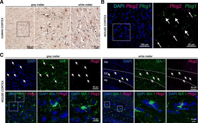
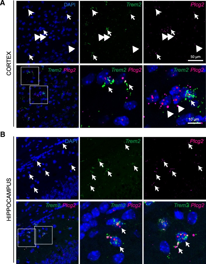
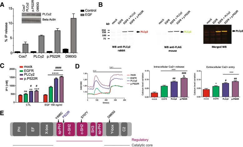
Methods: We assessed PLCG2 distribution in human and mouse brain tissue via immunohistochemistry and in situ hybridization. We transfected heterologous cell systems (COS7 and HEK293T cells) to determine the effect of the P522R AD-associated variant on enzymatic function using various orthogonal assays, including a radioactive assay, IP-One ELISA, and calcium assays.

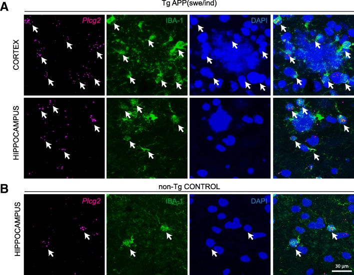
Results: PLCG2 expression is restricted primarily to microglia and granule cells of the dentate gyrus. Plcg2 mRNA is maintained in plaque-associated microglia in the cerebral tissue of an AD mouse model. Functional analysis of the p.P522R variant demonstrated a small hypermorphic effect of the mutation on enzyme function.

Conclusions: The PLCG2 P522R variant is protective against AD. We show that PLCG2 is expressed in brain microglia, and the p.P522R polymorphism weakly increases enzyme function. These data suggest that activation of PLCγ2 and not inhibition could be therapeutically beneficial in AD. PLCγ2 is therefore a potential target for modulating microglia function in AD, and a small molecule drug that weakly activates PLCγ2 may be one potential therapeutic approach.

Keywords: Dementia; Genetic variants; Immune response; Neuroinflammation; Phospholipase C.


Conflict of interest statement

Ethics approval and consent to participate

Work on human brain tissue was ethically approved by the Research Ethic Committee (08/H0718/54+5).

Animal procedures were approved by the University of Florida Institutional Animal Care and Use Committee.

Consent for publication

All the co-authors have given their consent for publication.

Competing interests

The authors declare that they have no competing interests.

Publisher’s Note

Springer Nature remains neutral with regard to jurisdictional claims in published maps and institutional affiliations.

Figures

Fig. 1
Fig. 1
PLCγ2 expression in the human and mouse cortical microglia. a IHC for PLCγ2 on the gray and white matter of the human prefrontal cortex. b Multiplex RNAScope for Plcg2 and Plcg1 on the adult mouse cortex. Small and big arrows point to RNASCope reaction for Plcg2 and Plcg1, respectively. c RNAScope for Plcg2 followed by IHC for IBA-1 on the gray and white matter of the adult mouse cortex. Arrows point to co-expression of the two markers. Ctx cortex, CC corpus callosum, Hp hippocampus
Fig. 2
Fig. 2
Plcg2 mRNA in microglia at amyloid plaques in a mouse model of AD. a RNAScope for Plcg2 in adult mouse neocortex and hippocampus of TgAPPswe/ind transgenic mice. Note expression of Plcg2 in IBA-1-labeled microglia surrounding the plaques (DAPI). Arrows point to co-labeling of IBA-1 and Plcg2. b RNAScope for Plcg2 in adult mouse hippocampus of a non-transgenic control mouse. Arrows point to co-labeling of IBA-1 and Plcg2
Fig. 3
Fig. 3
Plcg2 and Trem2 transcripts co-localize in the mouse brain. a Multiplex RNAScope for Plcg2 and Trem2 in the adult mouse cortex. Arrows point to co-expression, arrowheads indicate single-labeled cells. b Multiplex RNAScope for Plcg2 and Trem2 in the adult mouse hippocampal CA1. Arrows point to co-expression, arrowheads indicate single-labeled cells. Note co-localization in processes of putative microglia
Fig. 4
Fig. 4
PLCγ2 protective variant p.P552R shows a slightly hypermorphic activity by increasing intracellular calcium release. a PLC activity of the p.P522R variant under basal and stimulated conditions. Measurement of % IP release in non-transfected COS7 cells (COS7), COS7 cells transfected with pEGFPC1-PLCG2 constructs (common variant, PLCG2 WT; AD associated, rare variant PLCG2 P522R; and PLCG2 D993G (Ali5) mutation. Western blotting was used to confirm equal expression (anti-GFP, inset). SD is represented by error bars. The data are representative for 3 independent experiments. b HEK293T were transfected with mock, EGFR, EGFR and PLCG2 WT, or EGFR and PLCG2 P522R plasmids. c Quantification of IP1 by ELISA IP-One. Transfected HEK293T cells were stimulated with EGF 150 ng/ml and analyzed for IP1 quantification. ELISA reading were averaged of ± standard error (n = 3, *p < 0.05, **p < 0.01, ***p < 0.001, ****p < 0.0001, #p < 0.05, ####p < 0.0001, two-way ANOVA, Bonferroni multiple comparisons, *compared to control, #compared to EGFR). d Transfected HEK293T cells loaded with Fura-2AM were stimulated with human EGF 150 ng/ml. Intracellular calcium level was recorded by calculating the 340/380 nm excitation ratio at each 5 s. The net intracellular Ca2+ releases value was obtained by subtracting the average of 340/380 excitation ratio values between 0 and 50 s from the maximum 340/380 excitation ratio value between 60 and 200 s, after the EGF stimulation. The net extracellular Ca2+ entry value was obtained by subtracting the average 340/380 excitation ratio values between 285 and 380 s from the average of 340/380 excitation ratio values between 215 and 235 s after adding the extracellular calcium. Data in d are shown as averages of ± standard error (*p < 0.05, **p < 0.01, ***p < 0.001, #p < 0.05, ##p < 0.01, ###p < 0.001, & p < 0.05, one-way ANOVA, Tukey’s multiple comparison test, *compared to mock, #compared to EGFR, & compared to PLCγ2. The “n” is corresponding to an average of 45 reading from 3 independent transfection). e Linear representation of the domains of PLCG2 with locations of the point mutations (Y495C, Ali14; P522R, AD protective variant; S707Y, APLAID; D993G, Ali5)

References

    1. Gatz M, Reynolds CA, Fratiglioni L, Johansson B, Mortimer JA, Berg S, et al. Role of genes and environments for explaining Alzheimer disease. Arch Gen Psychiatry. 2006;63:168–174. doi: 10.1001/archpsyc.63.2.168. - DOI - PubMed
    1. Zhang B, Gaiteri C, Bodea LG, Wang Z, McElwee J, Podtelezhnikov AA, et al. Integrated systems approach identifies genetic nodes and networks in late-onset Alzheimer’s disease. Cell. 2013;153:707–720. doi: 10.1016/j.cell.2013.03.030. - DOI - PMC - PubMed
    1. Jones L, Lambert JC, Wang LS, Choi SH, Harold D, Vedernikov A, et al. Convergent genetic and expression data implicate immunity in Alzheimer’s disease. Alzheimers Dement. 2015;11:658–671. doi: 10.1016/j.jalz.2014.05.1757. - DOI - PMC - PubMed
    1. Efthymiou AG, Goate AM. Late onset Alzheimer’s disease genetics implicates microglial pathways in disease risk. Mol Neurodegener. 2017;12:43. doi: 10.1186/s13024-017-0184-x. - DOI - PMC - PubMed
    1. Guerreiro R, Wojtas A, Bras J, Carrasquillo M, Rogaeva E, Majounie E, et al. TREM2 variants in Alzheimer’s disease. N Engl J Med. 2013;368:117–127. doi: 10.1056/NEJMoa1211851. - DOI - PMC - PubMed

Publication types

Substances