. 2019 Apr 23:699:134-139.
doi: 10.1016/j.neulet.2019.01.056. Epub 2019 Jan 31.

Identification of β-synuclein on secretory granules in chromaffin cells and the effects of α- and β-synuclein on post-fusion BDNF discharge and fusion pore expansion

Affiliations

Identification of β-synuclein on secretory granules in chromaffin cells and the effects of α- and β-synuclein on post-fusion BDNF discharge and fusion pore expansion

Prabhodh S Abbineni et al. Neurosci Lett. .

Abstract

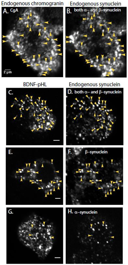
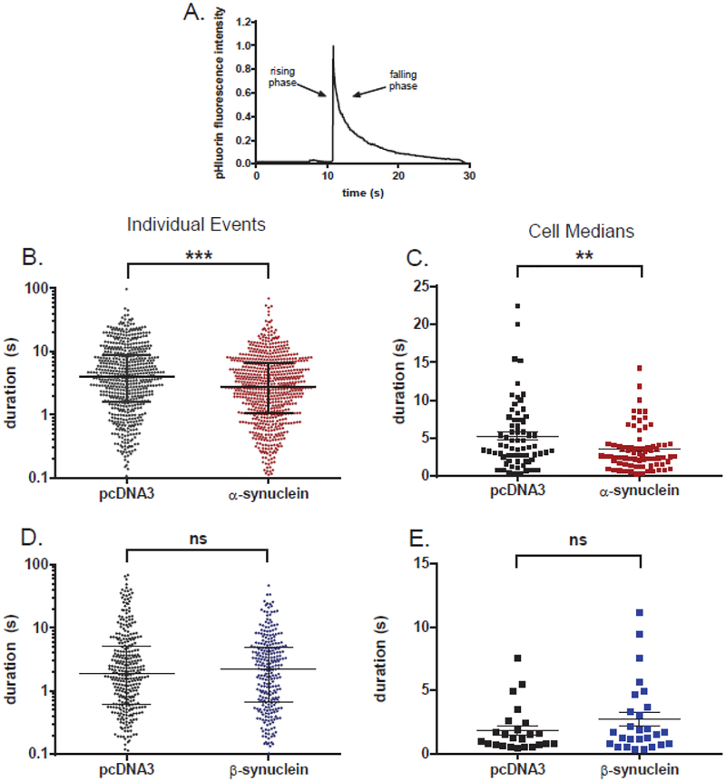
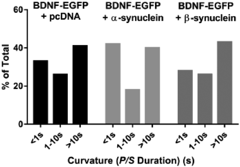
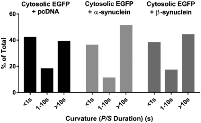
α-Synuclein is strongly implicated in the pathogenesis of Parkinson's disease as well as in other neurodegenerative diseases. However, its normal function in cells is not understood. The N-termini of α-, β-, and γ-synuclein contains six to seven 11-amino acid repeats that are predicted to form amphipathic helices. Membrane-binding and membrane-curving abilities of synuclein raise the possibility that synuclein could alter cellular processes that involve highly curved structures. In the present study we examined the localization of endogenous synuclein in bovine chromaffin cells by immunocytochemistry and its possible function to control protein discharge upon fusion of the granule with the plasma membrane by regulating the fusion pore. We found with quantitative immunocytochemistry that endogenous β-synuclein associates with secretory granules. Endogenous α-synuclein only rarely co-localizes with secretory granules. Overexpression of α-synuclein but not β-synuclein quickened the post- fusion discharge of BDNF-pHluorin by approxinately 30%. However, neither α- nor β-synuclein significantly altered curvature dynamics associated with fusion pore expansion that were measured by the combination of polarization and total internal reflection fluorescence microscopy (pTIRFM). Whatever the mechanism, the physiological significance of the small increased rate of post-fusion protein discharge caused by α-synuclein remains to be demonstrated, especially since endogenous β-, but not α-synuclein is the predominant synuclein isoform associated with chromaffin granules.

Keywords: Fusion pore; Immunocytochemistry; Secretion; Synuclein; TIRFM; pTIRFM.


Figures

Fig. 1.
Fig. 1.. Endogenous synuclein labels secretory granules in non-transfected cells (A, B) and newly synthesized chromaffin granules expressing exogenous BDNF-pHl in transfected cells (C-H).
Granules were identified with an antibody against chromogranin A (A) and endogenous synuclein with a monoclonal antibody that recognizes both the α- and β-isoforms of bovine synuclein (DSHB H3C, B). Immunocytochemistry was performed on chromaffin cells transiently expressing the chromaffin granule marker BDNF-pHl (C-H). (D) Endogenous synuclein was again detected by DSHB H3C, which recognizes both α- and β-synuclein. Arrowheads indicate some of the co-localized puncta. Synuclein puncta in a neighboring non-transfected cell are visible at the bottom of the image. (F) Endogenous synuclein as detected by an antibody specific for β-synuclein (Abcam 15532); (H) Endogenous synuclein as detected by an antibody specific for α-synuclein (Abcam 138501). Panels A and B, C and D, E and F and G and H are paired images of the same field of view. Note that panels C, E, and G show single transfected cells. Panels D, F, and H show several cells including the transfected cell. Scale bars = 2 μm.
Fig. 2.
Fig. 2.. α-Synuclein but not β-synuclein speeds the post-fusion discharge of BDNF-pHl.
(A) An example of the discharge of BDNF-pHl from a secretory granule upon fusion. (B) Scatter plot of event durations for α-synuclein-transfected cells and control cells (n=685 and 598 events, respectively). Note that the ordinate is log scale. ***, p = 0.0001 (Kolmogorov-Smirnov test). (C) Scatter plot of the median BDNF-pHl duration in each α-synuclein cell and control cell with greater than five events (n=73 and 67 cells, respectively). **, p = 0.006 (Student’s t-test). (D) Scatter plot of all event durations for β-synuclein-transfected and control cells (n=277 and 327 events, respectively). ns, non-significant difference (Kolmogorov-Smirnov test). (E) Scatter plot of the median BDNF-pHl duration in each β-synuclein-transfected and control cell with greater than five events (n=27 and 24 cells, respectively). ns, Student’s t-test. Horizontal bars indicate the median (longest bar) and the upper and lower quartiles in B and D. The mean of the cell medians (longest horizontal bar) and SEM (error bars) are shown in C and E.
Fig. 3.
Fig. 3.. Neither α-synuclein nor β-synuclein alters fusion pore curvature associated with BDNF discharge.
Fusion pore curvature associated with elevated K+-induced BDNF-EGFP discharge was monitored using pTIRF. The length of time P/S was elevated above the pre-fusion baseline was calculated for 88 BDNF-EGFP + pcDNA events, 88 BDNF + α-synuclein events, and 46 BDNF-EGFP + β-synuclein events and binned into three categories. The distributions were not significantly different.
Fig. 4.
Fig. 4.. Neither α-synuclein nor β-synuclein alters fusion pore curvature associated with fusion of granules not expressing exogenous protein.
Chromaffin cells were co-transfected with EGFP + pcDNA, EGFP + α-synuclein, or EGFP + β-synuclein. Upon stimulation, fusion of endogenous granules (i.e, not containing exogenous transfected protein) was identified as discrete, punctate changes in DiI fluorescence. At these sites, the length of time P/S remained elevated was measured for 88 EGFP + pcDNA events, 137 EGFP + α-synuclein events, 52 EGFP + β-synuclein events and binned into three categories. The distributions were not significantly different.

References

    1. Bendor JT, Logan TP, Edwards RH, The Function of alpha-Synuclein, Neuron 79 (2013) 1044–1066. - PMC - PubMed
    1. Larsen KE, Schmitz Y, Troyer MD, Mosharov E, Dietrich P, Quazi AZ, Savalle M, Nemani V, Chaudhry FA, Edwards RH, Stefanis L, Sulzer D, {alpha}-Synuclein Overexpression in PC12 and Chromaffin Cells Impairs Catecholamine Release by Interfering with a Late Step in Exocytosis, Journal of Neuroscience 26 (2006) 11915–11922. - PMC - PubMed
    1. Abeliovich A, Schmitz Y, Farinas I, Choi-Lundberg D, Ho WH, Castillo PE, Shinsky N, Verdugo JM, Armanini M, Ryan A, Hynes M, Phillips H, Sulzer D, Rosenthal A, Mice lacking alpha-synuclein display functional deficits in the nigrostriatal dopamine system, Neuron 25 (2000) 239–252. - PubMed
    1. Yavich L, Tanila H, Vepsalainen S, Jakala P, Role of alpha-synuclein in presynaptic dopamine recruitment, J Neurosci 24 (2004) 11165–11170. - PMC - PubMed
    1. Nemani VM, Lu W, Berge V, Nakamura K, Onoa B, Lee MK, Chaudhry FA, Nicoll RA, Edwards RH, Increased expression of alpha-synuclein reduces neurotransmitter release by inhibiting synaptic vesicle reclustering after endocytosis, Neuron 65 (2010) 66–79. - PMC - PubMed

Publication types