. 2019 Mar 15:367:51-61.
doi: 10.1016/j.taap.2019.01.024. Epub 2019 Feb 1.

Maternal titanium dioxide nanomaterial inhalation exposure compromises placental hemodynamics

Affiliations

Maternal titanium dioxide nanomaterial inhalation exposure compromises placental hemodynamics

Alaeddin B Abukabda et al. Toxicol Appl Pharmacol. .

Abstract

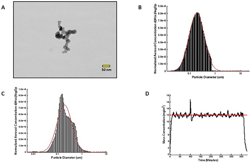
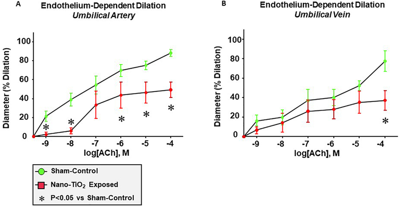
The fetal consequences of gestational engineered nanomaterial (ENM) exposure are unclear. The placenta is a barrier protecting the fetus and allowing transfer of substances from the maternal circulation. The purpose of this study was to determine the effects of maternal pulmonary titanium dioxide nanoparticle (nano-TiO2) exposure on the placenta and umbilical vascular reactivity. We hypothesized that pulmonary nano-TiO2 inhalation exposure increases placental vascular resistance and impairs umbilical vascular responsiveness. Pregnant Sprague-Dawley rats were exposed via whole-body inhalation to nano-TiO2 with an aerodynamic diameter of 188 ± 0.36 nm. On gestational day (GD) 11, rats began inhalation exposures (6 h/exposure). Daily lung deposition was 87.5 ± 2.7 μg. Animals were exposed for 6 days for a cumulative lung burden of 525 ± 16 μg. On GD 20, placentas, umbilical artery and vein were isolated, cannulated, and treated with acetylcholine (ACh), angiotensin II (ANGII), S-nitroso-N-acetyl-DL-penicillamine (SNAP), or calcium-free superfusate (Ca2+-free). Mean outflow pressure was measured in placental units. ACh increased outflow pressure to 53 ± 5 mmHg in sham-controls but only to 35 ± 4 mmHg in exposed subjects. ANGII decreased outflow pressure in placentas from exposed animals (17 ± 7 mmHg) compared to sham-controls (31 ± 6 mmHg). Ca2+-free superfusate yielded maximal outflow pressures in sham-control (63 ± 5 mmHg) and exposed (30 ± 10 mmHg) rats. Umbilical artery endothelium-dependent dilation was decreased in nano-TiO2 exposed fetuses (30 ± 9%) compared to sham-controls (58 ± 6%), but ANGII sensitivity was increased (-79 ± 20% vs -36 ± 10%). These results indicate that maternal gestational pulmonary nano-TiO2 exposure increases placental vascular resistance and impairs umbilical vascular reactivity.

Keywords: Engineered nanomaterials; Microcirculation; Placenta; Titanium dioxide nanoparticles.


Figures

Figure 1.
Figure 1.. Nano-TiO2 aerosol characterization.
(A) Transmission electron microscope image of a typical nano-TiO2 agglomerate generated with the high-pressure acoustical generator, (B) Size distribution of the nano-TiO2 aerosol (mobility diameter) sampled from the exposure chamber using a scanning mobility particle sizer (SMPS-light grey) and an aerodynamic particle sizer (APS-dark grey, negligible values). The red line represents a log-normal fit of the histogram (Count Median Diameter = 190 nm), (C) Size distribution of the nano-TiO2 aerosol (aerodynamic diameter) using a high resolution electrical low-pressure impactor (ELPI+). The red line represents a log-normal fit of the histogram (Count Median Diameter = 188 ± 0.36 nm). (D) Real-time mass concentration measurements of the nano-TiO2 aerosol during a typical inhalation exposure. The red line represents the target concentration, 12 mg/m3.
Figure 2.
Figure 2.. Ex vivo placental perfusion preparation.
(A) Umbilical artery (1); vein (2); placenta (3). (B) Input (arterial) perfusion pressure (1) is held constant while vasoactive agonists are added. Output pressure (2) and flow (3) (μl/min) are variables responsive to placental resistance.
Figure 3.
Figure 3.. Maternal nano-TiO2 inhalation exposure impairs endothelium-dependent placental hemodynamics.
Mean outflow pressures were measured in sham-control and nano-TiO2 exposed animals after the inflow pressure was increased in a stepwise manner and set at 0 mm Hg, 20 mm Hg, 40 mm Hg, 60 mm Hg and 80 mm Hg. Nano-TiO2 inhalation exposure altered placental hemodynamics and decreased outflow (venous) pressure in placentas in normal superfusate (A) and placentas treated with ACh (B) (n=8)., (P ≤ 0.05) * vs. sham-control group.
Figure 4.
Figure 4.. Maternal nano-TiO2 inhalation exposure does not affect endothelium-independent placental hemodynamics but increases ANGII sensitivity.
Mean outflow pressures were measured in sham-control and nano-TiO2 exposed animals after the inflow pressure was increased in a stepwise manner and set at 0 mm Hg, 20 mm Hg, 40 mm Hg, 60 mm Hg and 80 mm Hg. Maternal nano-TiO2 inhalation exposure did not affect endothelial-independent placental hemodynamics (A) but increased ANGII sensitivity (B) (n=8). , (P ≤ 0.05) * vs. sham-control group
Figure 5.
Figure 5.. Maternal nano-TiO2 inhalation impairs calcium-free placental hemodynamics.
Mean outflow pressures were measured in sham-control and nano-TiO2 exposed animals after the inflow pressure was increased in a stepwise manner and set at 0 mm Hg, 20 mm Hg, 40 mm Hg, 60 mm Hg and 80 mm Hg. Maternal nano-TiO2 inhalation exposure altered placental hemodynamics and decreased the outflow pressure in placentas placed in calcium-free superfusate (n=8)., (P ≤ 0.05) * vs. sham-control group
Figure 6.
Figure 6.. Maternal nano-TiO2 inhalation exposure and placental mean outflow pressure.
Mean outflow pressure at 80 mm Hg inflow pressure in both sham-control and nano-TiO2 exposed groups and all treatment conditions is shown (n=8)., (P ≤ 0.05) * vs. sham-control group
Figure 7.
Figure 7.. Maternal nano-TiO2 inhalation exposure impairs endothelium-dependent dilation of the umbilical artery and vein.
(A) Endothelium-dependent dilation of the umbilical artery, (B) endothelium-dependent dilation of the umbilical vein from sham-control and nano-TiO2 exposed animals was determined using pressure myography (n=12–14). Statistics were analyzed with two-way ANOVA., (P ≤ 0.05) * vs. sham-control group
Figure 8.
Figure 8.. Maternal nano-TiO2 inhalation exposure impairs endothelium-independent dilation of the umbilical artery and vein.
(A) Endothelium-independent dilation of the umbilical artery, (B) endothelium-independent dilation of the umbilical vein from sham-control and nano-TiO2 exposed animals was determined using pressure myography (n=12–14)., (P ≤ 0.05) * vs. sham-control group
Figure 9.
Figure 9.. Maternal nano-TiO2 inhalation exposure increases ANGII sensitivity of the umbilical artery.
(A) ANGII dose-response curve of the umbilical artery (A) and vein (B) from sham-control and nano-TiO2 exposed animals was determined using pressure myography (n=12–14)., (P ≤ 0.05) * vs. sham-control group
Figure 10.
Figure 10.. Placental immunohistochemistry.
Smooth muscle actin is green, Von-Willebrand Factor is red, and DAPI nuclear staining is blue. (A) Representative image showing immunohistochemistry of a placental section from maternal nano-TiO2 exposed animal. The purpose was to identify the microvascular architecture and the structural components of the placenta. (B) An enlarged (40×) image of a placental microvessel displaying an intact endothelium (green).

References

    1. Abukabda AB, Stapleton PA, McBride CR, Yi J, Nurkiewicz TR, 2017. Heterogeneous Vascular Bed Responses to Pulmonary Titanium Dioxide Nanoparticle Exposure. Front Cardiovasc Med 4,33. - PMC - PubMed
    1. Adamcakova-Dodd A, Monick MM, Powers LS, Gibson-Corley KN, Thorne PS, 2015. Effects of prenatal inhalation exposure to copper nanoparticles on murine dams and offspring. Part Fibre Toxicol 12,30. - PMC - PubMed
    1. Barker DJ 2007. The origins of the developmental origins theory. Journal of internal medicine, 261(5), 412–417. - PubMed
    1. Blum JL, Xiong JQ, Hoffman C, Zelikoff JT,2012. Cadmium associated with inhaled cadmium oxide nanoparticles impacts fetal and neonatal development and growth. Toxicol Sci. 126(2),478–86. - PMC - PubMed
    1. Ema M, Hougaard KS, Kishimoto A, Honda K, 2016. Reproductive and developmental toxicity of carbon-based nanomaterials: A literature review. Nanotoxicology. 10(4),391–412. - PubMed

Publication types