

Highly-branched poly(N-isopropyl acrylamide) functionalised with pendant Nile red and chain end vancomycin for the detection of Gram-positive bacteria
- PMID: 30711663
- PMCID: PMC6401204
- DOI: 10.1016/j.actbio.2019.01.066
Highly-branched poly(N-isopropyl acrylamide) functionalised with pendant Nile red and chain end vancomycin for the detection of Gram-positive bacteria
Abstract
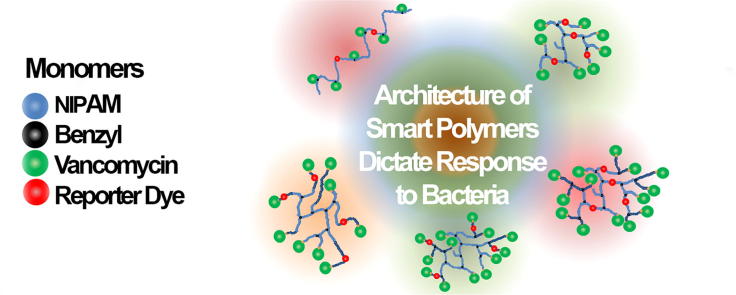
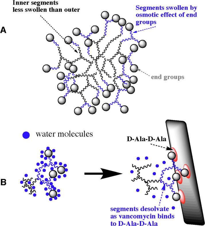
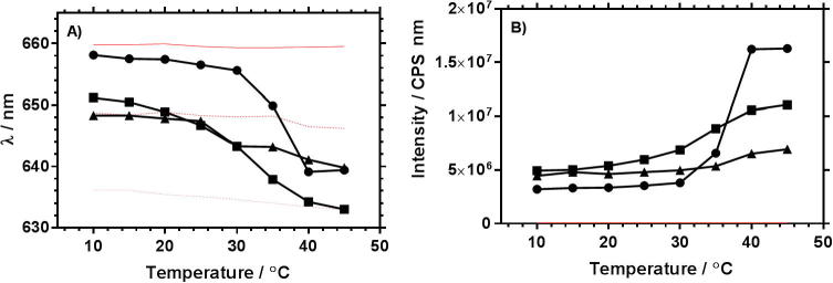
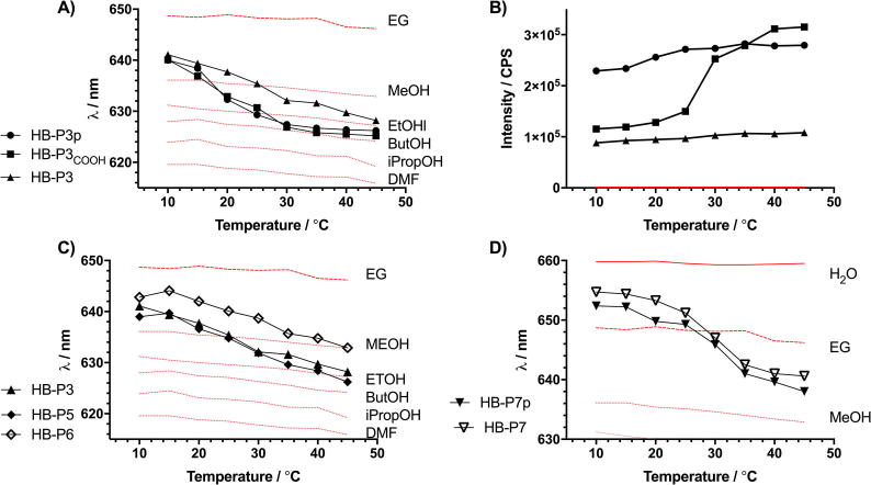
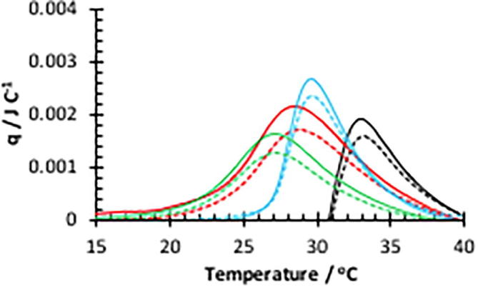
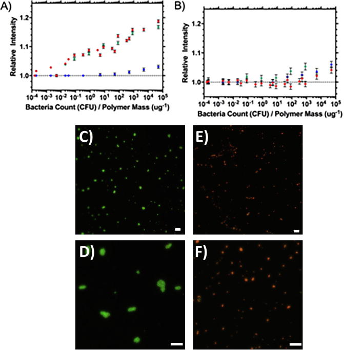
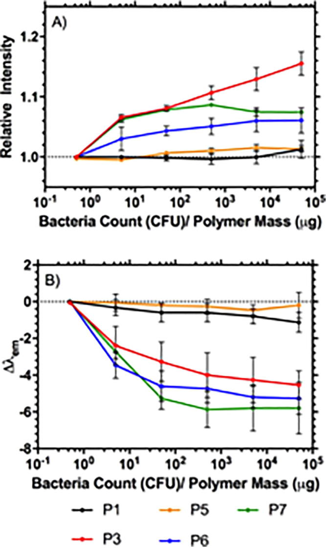
This study shows how highly branched poly(N-isopropyl acrylamide) (HB-PNIPAM) with a chain pendant solvatochromic dye (Nile red) could provide a fluorescence signal, as end groups bind to bacteria and chain segments become desolvated, indicating the presence of bacteria. Vancomycin was attached to chain ends of HB-PNIPAM or as pendant groups on linear polymers each containing Nile red. Location of the dye was varied between placement in the core of the branched polymer coil or the outer domains. Both calorimetric and fluorescence data showed that branched polymers responded to binding of both the peptide target (D-Ala-D-Aa) and bacteria in a different manner than analogous linear polymers; binding and response was more extensive in the branched variant. The fluorescence data showed that only segments located in the outer domains of branched polymers responded to binding of Gram-positive bacteria with little response when linear analogous polymer or branched polymer with the dye in the inner core was exposed to Staphylococcus aureus.
Keywords: Bacterial sensor; Diagnostic device; Polymer architecture; Solvatochromism; Specificity; Stimuli responsive.
Copyright © 2019 Acta Materialia Inc. Published by Elsevier Ltd. All rights reserved.
Figures
References
-
- Shepherd J., Sarker P., Swindells K., Douglas I., MacNeil S., Swanson L., Rimmer S. Binding bacteria to highly branched poly(N-isopropyl acrylamide) modified with vancomycin induces the coil-to-globule transition. J. Am. Chem. Soc. 2010;132(6):1736–1737. - PubMed
-
- Sarker P., Shepherd J., Swindells K., Douglas I., MacNeil S., Swanson L., Rimmer S. Highly branched polymers with polymyxin end groups responsive to Pseudomonas aeruginosa. Biomacromolecules. 2011;12(1):1–5. - PubMed
-
- Plenderleith R.A., Pateman C.J., Rodenburg C., Haycock J.W., Claeyssens F., Sammon C., Rimmer S. Arginine-glycine-aspartic acid functional branched semi-interpenetrating hydrogels. Soft Matter. 2015;11(38):7567–7578. - PubMed
-
- Zhang Y., Furyk S., Bergbreiter D.E., Cremer P.S. Specific ion effects on the water solubility of macromolecules: PNIPAM and the hofmeister series. J. Am. Chem. Soc. 2005;127(41):14505–14510. - PubMed
-
- Sakota K., Tabata D., Sekiya H. Macromolecular crowding modifies the impact of specific hofmeister ions on the coil-globule transition of PNIPAM. J. Phys. Chem. B. 2015;119(32):10334–10340. - PubMed
Publication types
MeSH terms
Substances
Grants and funding
LinkOut - more resources
Full Text Sources
Other Literature Sources
Molecular Biology Databases
