

Mapping acute lesion locations to physiological swallow impairments after stroke
- PMID: 30711683
- PMCID: PMC6357850
- DOI: 10.1016/j.nicl.2019.101685
Mapping acute lesion locations to physiological swallow impairments after stroke
Abstract
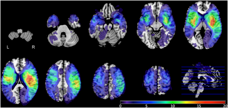
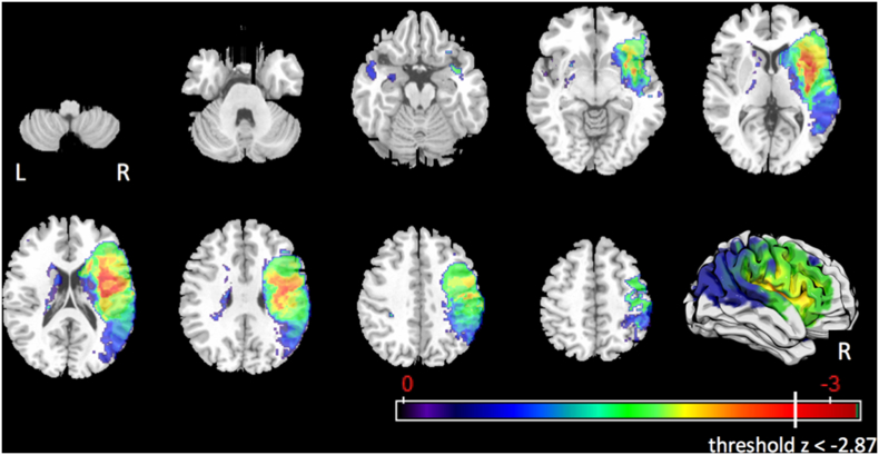
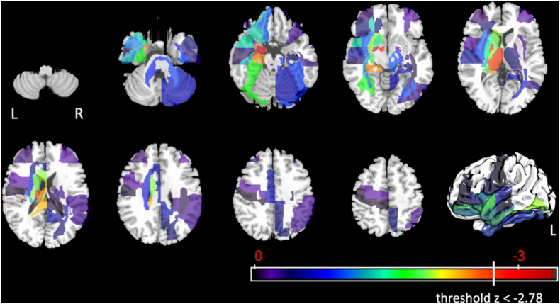
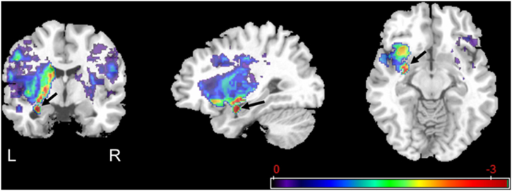
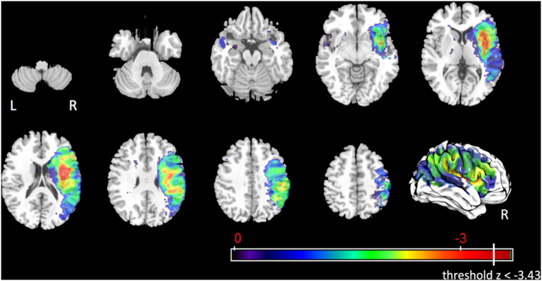
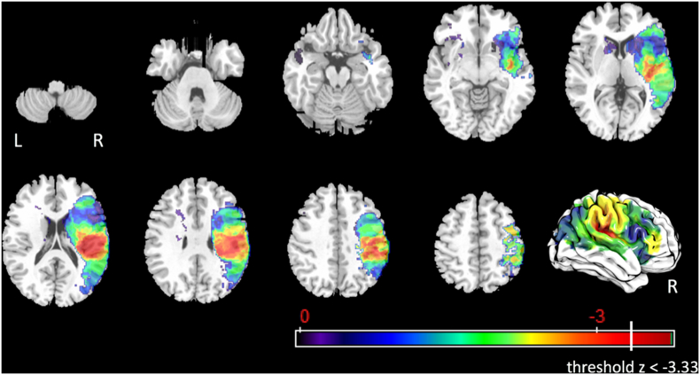
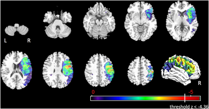
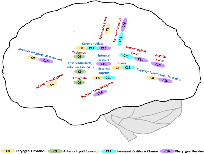
Dysphagia is a common deficit after a stroke, and it is frequently associated with pneumonia, malnutrition, dehydration, and poor quality of life. It is not yet fully clear which brain regions are directly related to swallowing, and how lesions affect swallow physiology. This study aimed to assess the statistical relationship between acute stroke lesion locations and impairment of specific aspects of swallow physiology. We performed lesion symptom mapping with 68 retrospectively recruited, acute, first-ever ischemic stroke patients. Lesions were determined on diffusion weighted MRI scans. Post-stroke swallow physiology was determined using the Modified Barium Swallow Study Impairment Profile (MBSImP©™). The relationship between brain lesion location and 17 physiological aspects of swallowing were tested using voxel-based and region-based statistical associations corrected for multiple comparisons using permutation thresholding. We found that laryngeal elevation, anterior hyoid excursion, laryngeal vestibular closure, and pharyngeal residue were associated with lesioned voxels or regions of interests. All components showed distinct and overlapping lesion locations, mostly in the right hemisphere, and including cortical regions (inferior frontal gyrus, pre- and postcentral gyrus, supramarginal gyrus, angular gyrus, superior temporal gyrus, insula), subcortical regions (thalamus, amygdala) and white matter tracts (superior longitudinal fasciculus, corona radiata, internal capsule, external capsule, ansa lenticularis, lenticular fasciculus). Our findings indicate that different aspects of post-stroke swallow physiology are associated with distinct lesion locations, primarily in the right hemisphere, and primarily including sensory-motor integration areas and their corresponding white matter tracts. Future studies are needed to expand on our findings and thus, support the development of a neuroanatomical model of post-stroke swallow physiology and treatment approaches targeting the neurophysiological underpinnings of swallowing post stroke.
Keywords: Deglutition; Deglutition disorders; Lesion analysis; Magnetic resonance imaging; Stroke.
Copyright © 2019 The Authors. Published by Elsevier Inc. All rights reserved.
Figures
References
-
- Ashburner J., Friston K.J. Unified segmentation. NeuroImage. 2005;26:839–851. - PubMed
-
- Augustine J.R. Circuitry and functional aspects of the insular lobe in primates including humans. Brain Res. Brain Res. Rev. 1996;22:229–244. - PubMed
-
- Barikroo A., Carnaby G., Crary M. Effects of age and bolus volume on velocity of hyolaryngeal excursion in healthy adults. Dysphagia. 2015;30:558–564. - PubMed
-
- Benjamin E.J., Blaha M.J., Chiuve S.E., Cushman M., Das S.R., Deo R., de Ferranti S.D., Floyd J., Fornage M., Gillespie C., Isasi C.R., Jimenez M.C., Jordan L.C., Judd S.E., Lackland D., Lichtman J.H., Lisabeth L., Liu S., Longenecker C.T., Mackey R.H., Matsushita K., Mozaffarian D., Mussolino M.E., Nasir K., Neumar R.W., Palaniappan L., Pandey D.K., Thiagarajan R.R., Reeves M.J., Ritchey M., Rodriguez C.J., Roth G.A., Rosamond W.D., Sasson C., Towfighi A., Tsao C.W., Turner M.B., Virani S.S., Voeks J.H., Willey J.Z., Wilkins J.T., Wu J.H., Alger H.M., Wong S.S., Muntner P. Heart disease and stroke statistics-2017 update: a report from the American Heart Association. Circulation. 2017;135:e146–e603. - PMC - PubMed
Publication types
MeSH terms
Grants and funding
LinkOut - more resources
Full Text Sources
Medical
