

Massive generalized crystal-storing histiocytosis associated with extracellular crystalline nephropathy: clinical, immunohistochemical, and ultrastructural studies of a unique disorder and review of the literature
- PMID: 30712253
- PMCID: PMC6620229
- DOI: 10.1007/s13730-019-00385-5
Massive generalized crystal-storing histiocytosis associated with extracellular crystalline nephropathy: clinical, immunohistochemical, and ultrastructural studies of a unique disorder and review of the literature
Abstract
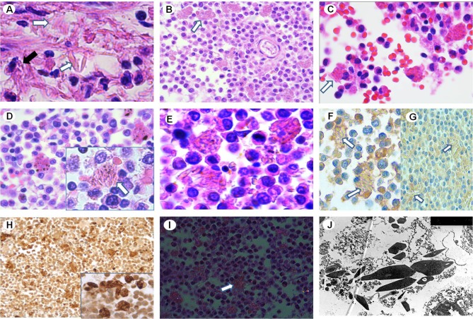
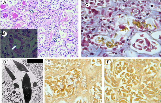
Crystal-storing histiocytosis (CSH) is a rare disorder characterized by the accumulation of nonneoplastic histiocytes containing intracytoplasmic crystallized immunoglobulins. In most cases, there is an associated underlying lymphoplasmacytic neoplasm expressing Ig kappa light chain. About 131 cases of CSH have been identified. There is a localized and a generalized form of CSH and it can involve several sites including bone marrow, lungs, lymph nodes, liver, spleen, gastrointestinal tract, and kidney. Generalized CSH is less frequent and involves multiple organs and tends to have a worst prognosis than localized CSH. Around 20 cases of renal involvement in CSH have been reported so far. Paraprotein-induced crystalline nephropathy can be divided into two categories based on whether the crystals in the kidney are intracellular (including light chain proximal tubulopathy with crystals and CSH) or extracellular (including the crystalline variant of myeloma cast nephropathy and crystalglobulin-induced nephropathy). The former tends to present with slowly worsening kidney dysfunction and generally has a good prognosis, whereas the latter usually presents with rapidly progressive renal failure and is associated with poor renal outcome. We present a case of generalized CSH associated with extracellular crystalline nephropathy with a fulminant and fatal clinical course. Kappa light-chain crystals were found exclusively extracellularly within the tubular lumen, not within the tubular epithelial cells nor the histiocytes, and the massive presence of those precipitates led to the acute renal failure. Consequently, we review the renal involvement in CSH in the literature and discuss the unique mechanism of renal injury in this case.
Keywords: Crystal-storing histiocytosis; Crystalline nephropathy; Histiocytes; Kappa light chains; Kidney.
Conflict of interest statement
The authors have declared that no conflict of interest exists.
Figures

References
-
- Kanagal-Shamanna R, Xu-Monette ZY, Miranda RN, Dogan A, Zou D, Luthra R, Weber DM, O’Malley DP, Jorgensen JL, Khoury JD, Bueso-Ramos CE, Orlowski RZ, Medeiros LJ, Young KH. Crystal-storing histiocytosis: a clinicopathological study of 13 cases. Histopathology. 2016;68:482–491. doi: 10.1111/his.12768. - DOI - PMC - PubMed
Publication types
MeSH terms
LinkOut - more resources
Full Text Sources
Medical
Miscellaneous
