

Zika Virus Protease Cleavage of Host Protein Septin-2 Mediates Mitotic Defects in Neural Progenitors
- PMID: 30713029
- PMCID: PMC6690588
- DOI: 10.1016/j.neuron.2019.01.010
Zika Virus Protease Cleavage of Host Protein Septin-2 Mediates Mitotic Defects in Neural Progenitors
Abstract
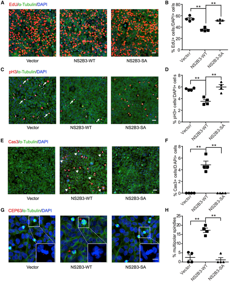
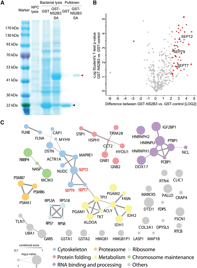
Zika virus (ZIKV) targets neural progenitor cells in the brain, attenuates cell proliferation, and leads to cell death. Here, we describe a role for the ZIKV protease NS2B-NS3 heterodimer in mediating neurotoxicity through cleavage of a host protein required for neurogenesis. Similar to ZIKV infection, NS2B-NS3 expression led to cytokinesis defects and cell death in a protease activity-dependent fashion. Among binding partners, NS2B-NS3 cleaved Septin-2, a cytoskeletal factor involved in cytokinesis. Cleavage of Septin-2 occurred at residue 306 and forced expression of a non-cleavable Septin-2 restored cytokinesis, suggesting a direct mechanism of ZIKV-induced neural toxicity. VIDEO ABSTRACT.
Keywords: Zika; activated caspase; cytokinesis; microcephaly; protease; septin.
Copyright © 2019. Published by Elsevier Inc.
Conflict of interest statement
DECLARATION OF INTERESTS
The authors declare no competing interests.
Figures


Comment in
-
Dissecting the Toxic Effects of Zika Virus Proteins on Neural Progenitor Cells.Neuron. 2019 Mar 20;101(6):989-991. doi: 10.1016/j.neuron.2019.03.009. Neuron. 2019. PMID: 30897362
Similar articles
-
Dissecting the Toxic Effects of Zika Virus Proteins on Neural Progenitor Cells.Neuron. 2019 Mar 20;101(6):989-991. doi: 10.1016/j.neuron.2019.03.009. Neuron. 2019. PMID: 30897362
-
Analysis of cellular proteome changes in response to ZIKV NS2B-NS3 protease expression.Biochim Biophys Acta Proteins Proteom. 2019 Feb;1867(2):89-97. doi: 10.1016/j.bbapap.2018.10.016. Epub 2018 Oct 31. Biochim Biophys Acta Proteins Proteom. 2019. PMID: 30391636
-
Zika NS2B is a crucial factor recruiting NS3 to the ER and activating its protease activity.Virus Res. 2020 Jan 2;275:197793. doi: 10.1016/j.virusres.2019.197793. Epub 2019 Oct 29. Virus Res. 2020. PMID: 31676367
-
The Structure of the Zika Virus Protease, NS2B/NS3pro.Adv Exp Med Biol. 2018;1062:131-145. doi: 10.1007/978-981-10-8727-1_10. Adv Exp Med Biol. 2018. PMID: 29845530 Review.
-
NS2B-NS3 protease inhibitors as promising compounds in the development of antivirals against Zika virus: A systematic review.J Med Virol. 2022 Feb;94(2):442-453. doi: 10.1002/jmv.27386. Epub 2021 Oct 20. J Med Virol. 2022. PMID: 34636434
Cited by
-
TRIM5α: A Protean Architect of Viral Recognition and Innate Immunity.Viruses. 2024 Jun 21;16(7):997. doi: 10.3390/v16070997. Viruses. 2024. PMID: 39066160 Free PMC article. Review.
-
The RNA polymerase of cytoplasmically replicating Zika virus binds with chromatin DNA in nuclei and regulates host gene transcription.Proc Natl Acad Sci U S A. 2022 Dec 6;119(49):e2205013119. doi: 10.1073/pnas.2205013119. Epub 2022 Nov 29. Proc Natl Acad Sci U S A. 2022. PMID: 36442102 Free PMC article.
-
ZIKV viral proteins and their roles in virus-host interactions.Sci China Life Sci. 2021 May;64(5):709-719. doi: 10.1007/s11427-020-1818-4. Epub 2020 Oct 14. Sci China Life Sci. 2021. PMID: 33068285 Free PMC article. Review.
-
Inter-organelle interactions between the ER and mitotic spindle facilitates Zika protease cleavage of human Kinesin-5 and results in mitotic defects.iScience. 2021 Mar 31;24(5):102385. doi: 10.1016/j.isci.2021.102385. eCollection 2021 May 21. iScience. 2021. PMID: 33997675 Free PMC article.
-
An overview of Zika virus genotypes and their infectivity.Rev Soc Bras Med Trop. 2022 Sep 30;55:e02632022. doi: 10.1590/0037-8682-0263-2022. eCollection 2022. Rev Soc Bras Med Trop. 2022. PMID: 36197380 Free PMC article. Review.
References
-
- Beites CL, Xie H, Bowser R, andTrimble WS (1999). The septin CDCrel-1 binds syntaxin and inhibits exocytosis. Nat. Neurosci. 2, 434–439. - PubMed
-
- Besnard M, Eyrolle-Guignot D, Guillemette-Artur P, Lastère S, Bost-Bezeaud F, Marcelis L, Abadie V, Garel C, Moutard ML, Jouannic JM, et al. (2016). Congenital cerebral malformations and dysfunction in fetuses and newborns following the 2013 to 2014 Zika virus epidemic in French Polynesia. Euro Surveill. 21, 13. - PubMed
-
- Butler D (2016). Zika virus: Brazil’s surge in small-headed babies questioned by report. Nature 530, 13–14. - PubMed
Publication types
MeSH terms
Substances
Grants and funding
LinkOut - more resources
Full Text Sources
Other Literature Sources
Medical
Research Materials
