. 2019 Mar 6;27(3):661-672.
doi: 10.1016/j.ymthe.2019.01.011. Epub 2019 Jan 22.

Endogenously Expressed Antigens Bind Mammalian RNA via Cationic Domains that Enhance Priming of Effector CD8 T Cells by DNA Vaccination

Affiliations

Endogenously Expressed Antigens Bind Mammalian RNA via Cationic Domains that Enhance Priming of Effector CD8 T Cells by DNA Vaccination

Jana Krieger et al. Mol Ther. .

Abstract

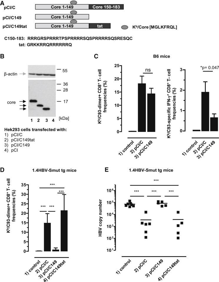
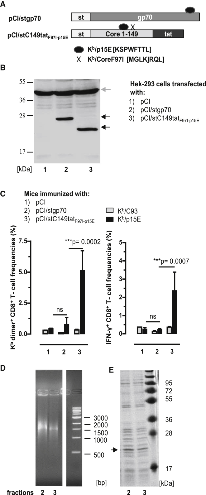
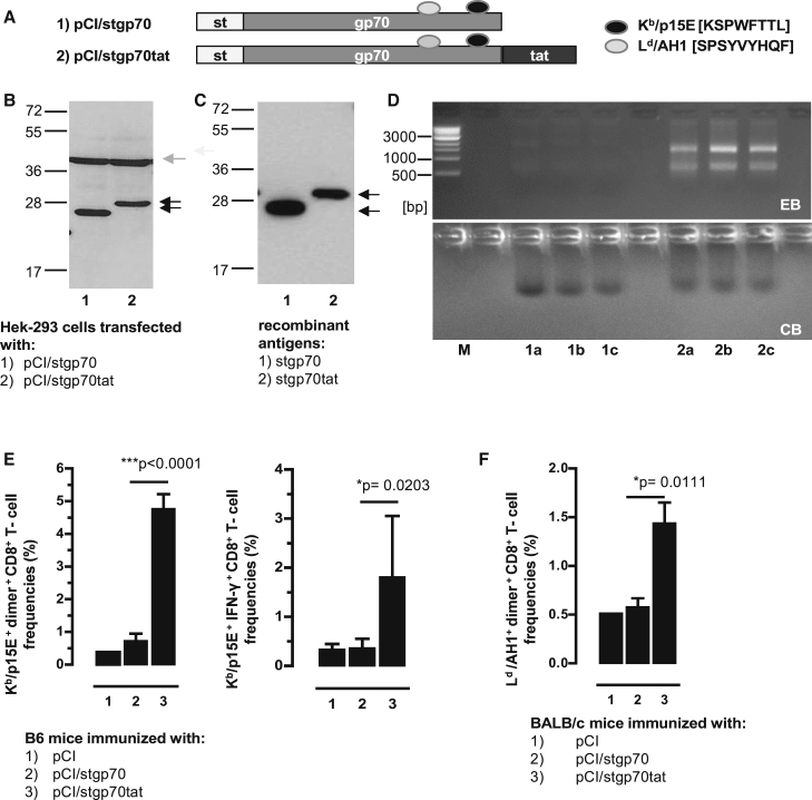
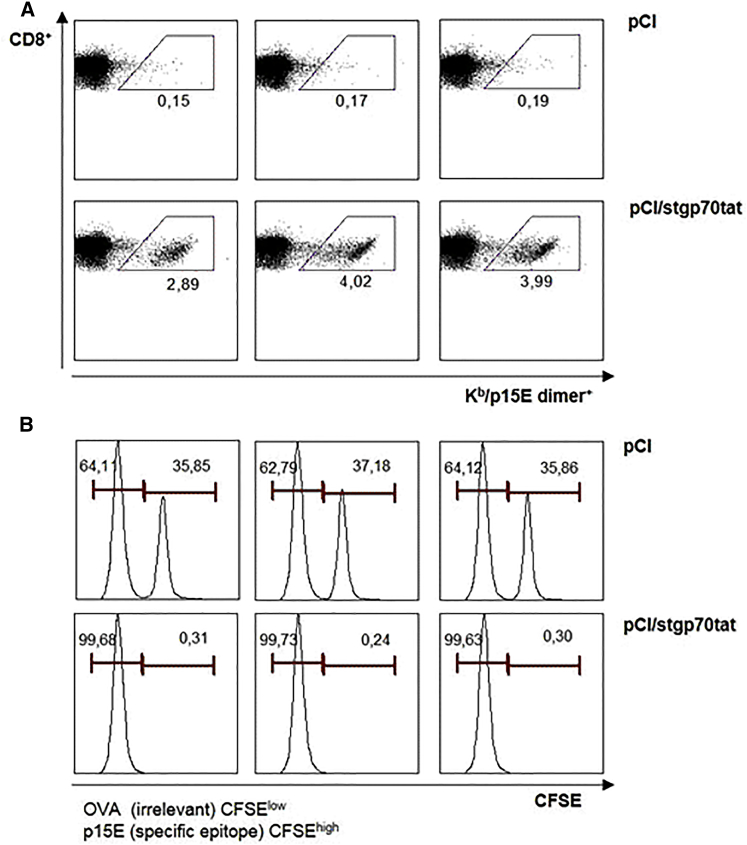
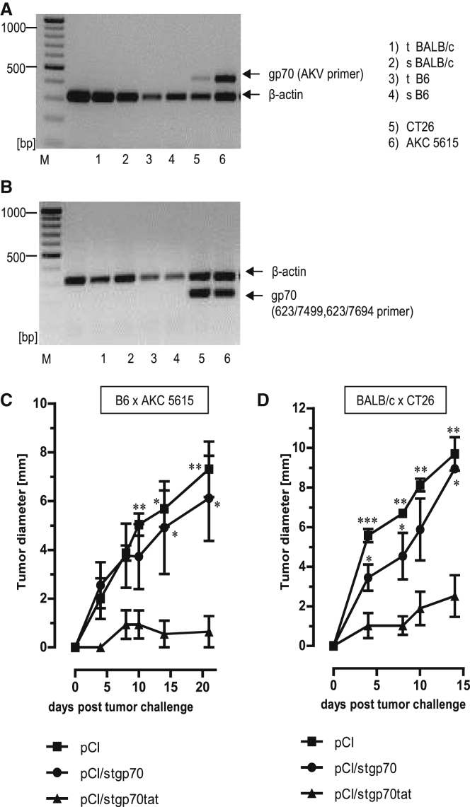
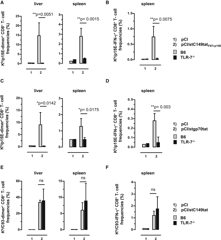
Hepatitis B virus (HBV) core (HBV-C) antigens with homologous or heterologous HIV-tat48-57-like (HBV-C149tat) cationic domains non-specifically bind cellular RNA in vector-transfected cells. Here, we investigated whether RNA-binding to cationic domains influences the immunogenicity of endogenously expressed antigens delivered by DNA vaccination. We initially evaluated induction of HBV-C (Kb/C93)-specific CD8+ T cell responses in C57BL/6J (B6) and 1.4HBV-Smut transgenic (tg) mice that harbor a replicating HBV genome in hepatocytes by DNA immunization. RNA-binding HBV-C and HBV-C149tat antigens moderately enhanced Kb/C93-specific CD8+ T cells in B6 mice as compared with RNA-free HBV-C149 antigen (lacking cationic domains). However, only the RNA-binding antigens elicited Kb/C93-specific CD8+ T cells that inhibited HBV replication in 1.4HBV-Smut tg mice. Moreover, RNA-binding to designer antigens, which express a Kb/p15E epitope from an endogenous murine leukemia virus-derived tumor-specific gp70 protein, was crucial to prime tumor-rejecting effector CD8+ T cells in B6 mice. Antigen-bound endogenous RNAs function as a Toll-like receptor 7 (TLR-7) ligand and stimulated priming of Kb/p15E-specific CD8+ T cells in B6, but not TLR-7-/-, mice. Antigen-bound cellular RNAs thus function as an endogenous natural adjuvant in in vivo vector-transfected cells, and thus are an attractive tool to induce and/or enhance effector CD8+ T cell responses directed against chronic viral infections or tumor self-antigens by DNA vaccination.

Keywords: CD8 T cells; DNA vaccination; RNA-binding; adjuvant; cationic domains.


Figures

Figure 1
Figure 1
Induction of Kb/C93-Specific CD8+ T Cell Responses by DNA-Based Vaccines (A) Schematic presentation of HBV-C, HBV-C149, and HBV-C149tat antigens. The position and amino acid (aa) sequences of the Kb/C93-100 epitope, the C150–183, and HIV-tat-derived cationic domains are given. (B) HEK293 cells were transiently transfected with the indicated plasmids, lysed, and processed for SDS-PAGE followed by core- and actin-specific western blot analyses. The positions of β-actin (gray arrow), HBV core antigens (black arrows), and a molecular weight marker (in kDa) are indicated. This image was generated from two films with different exposure times (1 min for the lower part and 3 min for the upper part). (C) B6 mice were either non-treated (group 1; n = 4) or injected with pCI/C (group 2; n = 6) and pCI/C149 (group 3; n = 6). Twelve days post-immunization, Kb/C93-specific dimer+ CD8+ T cell responses were determined in the liver. The mean percentage of Kb/C93-specific dimer+ CD8+ T cells in the hepatic CD8+ T cell populations (±SD) is shown (left panel). Furthermore, spleen cells were ex vivo re-stimulated with Kb/C93 or an irrelevant Kb/Ova257-264 peptide. Specific IFN-γ+ CD8+ T cell frequencies were determined by FCM, and Kb/Ova257-specific background values were subtracted. The mean percentage of Kb/C93-specific IFN-γ+ CD8+ T cells in the splenic CD8+ T cell population (±SD) is shown (right panel). The statistical significance of differences in dimer+ or IFN-γ+ CD8+ T cell frequencies between groups 2 and 3 was determined by the unpaired Student’s t test. *p values <0.05 were considered statistically significant. (D and E) 1.4HBV-Smut tg mice (four to six mice per group) were either non-treated (group 1) or injected with pCI/C (group 2), pCI/C149 (group 3), and pCI/C149tat (group 4) DNA. (D) Twelve days post-immunization, Kb/C93-specific dimer+ CD8+ T cell responses were determined in the liver. The mean percentage of Kb/C93-specific dimer+ CD8+ T cells in the hepatic CD8+ T cell populations (±SD) is shown. (E) HBV replication was determined in the liver of non-treated (control) and vaccinated 1.4HBV-Smut tg mice by real-time qPCR as described in the Materials and Methods. (D and E) The statistical significance of differences between different groups of 1.4HBV-Smut tg mice was determined by one-way ANOVA followed by Tukey’s multiple comparison test. *p < 0.05 and ***p < 0.001 were considered significant. ns, not significant.
Figure 2
Figure 2
Induction of Kb/p15E-Specific CD8+ T Cell Responses by DNA Vaccination (A) Schematic presentation of pCI/stgp70 and pCI/stC149tatF97I-p15E antigens. The pCI/stgp70 vector is composed of an NH2-terminal strep-tag (st) and a 289-residue gp70327–615 fragment. PCI/stC149tatF97I-p15E encodes for the HBV-stC149tat antigen with the Kb/p15E sequence cloned into the major immunodominant region. Additionally, it harbors a point mutation at position 97 in the HBV-C encoding region, silencing the Kb/C93 epitope. The corresponding Kb/C93F97I sequence is indicated by an X. (B) Lysates of HEK293 cells transiently transfected with pCI/stgp70 and pCI/stC149tatF97I-p15E were processed for SDS-PAGE followed by strep-tag- (black arrows) and actin-specific (gray arrow) western blot analyses. The position of the molecular weight marker (in kDa) is indicated. (C) B6 mice were immunized intramuscularly (i.m.) with pCI (group 1; n = 3), pCI/stgp70 (group 2; n = 6), and pCI/stC149tatF97I-p15E (group 3; n = 4). Twelve days post-immunization, antigen-specific (Kb/p15E and Kb/C93) dimer+ and IFN-γ+ CD8+ T cell frequencies were determined in the spleen by FCM. The mean percentages of dimer+ and IFN-γ+ CD8+ T cells in the splenic CD8+ T cell populations (+SD) are shown. The statistical significance of differences in Kb/p15E-specific frequencies between indicated groups was determined by the unpaired Student’s t test. *p < 0.05, **p < 0.01, and ***p < 0.001 were considered statistically significant. (D and E) HEK293 cells were transiently transfected with the pCI/stC149tatF97I-p15E. Recombinant stCtatF97I-p15E antigen was purified from cell lysates, and elution fractions 2 and 3 were analyzed on a native agarose gel stained with ethidium bromide (EB) (D) and on an SDS-containing polyacrylamide gel stained with coomassie blue (CB) (E). The molecular weight marker (in kDa) and DNA marker (in bp) are shown. ns, not significant.
Figure 3
Figure 3
RNA-Binding of the stgp70tat Fusion Protein Facilitated Priming of gp70-Specific CD8+ T Cell Responses (A) Schematic presentation of stgp70 and stgp70tat antigens. The positions of the strep-tag (st), the gp70327–615 fragment, and the HIV-tat-like cationic domain, as well as the Ld and Kb binding epitopes, are indicated. (B) Same numbers of HEK293 cells were transiently transfected with the pCI/stgp70 and pCI/stgp70tat vectors. Forty-eight hours post-transfection, cell lysates were processed for SDS-PAGE followed by strep-tag and β-actin-specific western blot. The position of the antigens (black arrows), β-actin (gray arrow), and the molecular weight marker (in kDa) are indicated. (C) Recombinant proteins were purified from lysates of the same numbers of transfected HEK293 cells as described in the Materials and Methods. Samples of pooled elution fractions (see D, lanes 1a, 1b, 1c and 2a, 2b, and 2c) were processed for SDS-PAGE, and antigens were detected by strep-tag-specific western blot. (D) Elution fractions of purified stgp70 (lanes 1a, 1b, and 1c) and stgp70tat (lanes 2a, 2b, and 2c) were analyzed by native agarose gel electrophoresis followed by EB (upper panel) and subsequent CB staining of the gel (lower panel). (E and F) B6 mice (five to seven mice per group) (E) and BALB/c mice (four mice per group) (F) were vaccinated with pCI (group 1), pCI/stgp70 (group 2), and pCI/stgp70tat (group 3) vectors. Kb/p15E-specific dimer+ and IFN-γ+ CD8+ T cells (E) and Ld/AH1-specific dimer+ CD8+ T cells (F) in the spleen were determined 12 days post-injection by FCM. (E and F) The mean percentages of dimer+ or IFN-γ+ CD8+ T cells in the splenic CD8+ T cell populations (+SD) are shown. The statistical significance of differences between groups 2 and 3 were determined by the unpaired Student’s t test. *p < 0.05 and ***p < 0.001 were considered statistically significant.
Figure 4
Figure 4
Antigen-Specific In Vivo Killing of Peptide-Pulsed Target Cells Groups of B6 mice (n = 3) were vaccinated with pCI or pCI/stgp70tat vectors. Twelve days post-injection, peptide-pulsed and CFSE-labeled (irrelevant Kb/Oav257 peptide/low CFSE and Kb/p15E peptide/high CFSE) naive splenocytes of non-treated B6 mice were mixed and adoptively transferred (i.v.) into vaccinated recipients. Spleens were harvested 16 h post-transfer and analyzed for antigen-specific p15E dimer+ CD8+ T cells (A), and CSFE-labeled cells were quantified by FCM (B).
Figure 5
Figure 5
Rejection of gp70-Expressing CT26 and AKC-5615 Tumors in Vaccinated B6 and BALB/c Mice (A and B) RNA was purified from thymus (t) and spleen (s) of B6 and BALB/c mice or CT26 and AKC-5615 cancer cell lines and transcribed into cDNA followed by PCR with gp70-specific AKV primers (A), 623/7499, 623/7694 primers (B), as well as β-actin-specific primers as described in the Materials and Methods. PCR products were analyzed on 1.5% agarose gels stained with EB. Positions of β-actin- and gp70-specific PCR products are indicated. (C) Growth of AKC-5615 tumors in B6 mice vaccinated with pCI/stgp70, pCI/stgp70tat, or pCI. Five mice per group were vaccinated with the indicated vectors, and 5 × 105 AKC-5615 cells were transplanted subcutaneously (s.c.) at 12 days post-vaccination. (D) Growth of CT26 tumors in BALB/c mice vaccinated with pCI/stgp70, pCI/stgp70tat, or pCI. Five mice per group were vaccinated and boosted (3 weeks after the first injection) with the indicated vectors. Mice were transplanted s.c. with 106 CT26 cells at day 12 after the second DNA injection. (C and D) Tumor growth was followed daily by serial measurements of tumor size at two perpendicular diameters. Mean values of five mice per group ± SEM are shown. The statistical significance of differences in tumor size between groups at the indicated time points was determined by one-way ANOVA followed by Tukey’s multiple comparison test. *p < 0.05, **p < 0.01, and ***p < 0.001 were considered statistically significant.
Figure 6
Figure 6
CD8+ T Cell Induction in Vaccinated B6 and TLR7−/− Mice TLR7−/− and B6 mice were immunized intramuscularly (i.m.) (n = 3–4 mice/group) with empty pCI and pCI/stC149tatF97I-p15E (A and B), with empty pCI and pCI/stgp70tat (C and D), or with empty pCI and pCI/stC149tat (E and F). Twelve days post-injection, antigen-specific dimer+ CD8+ T cell responses were determined in the liver and spleen. Furthermore, spleen cells were ex vivo re-stimulated with Kb/C93 and Kb/p15E peptides or an irrelevant Kb/Ova257 peptide. Specific IFN-γ+ CD8+ T cell frequencies were determined by FCM, and Kb/Ova257-specific background values were subtracted. The mean percentages of dimer+ (A and C) and IFN-γ+ Kb/p15E-specific CD8+ T cells (B and D) in the corresponding CD8+ T cell populations (+SD) are shown. Furthermore, the mean percentages of dimer+ (E) and IFN-γ+ Kb/C93-specific CD8+ T cells (F) in the corresponding CD8+ T cell populations (+SD) are shown. The statistical significance of differences between vaccinated B6 and TLR7−/− mice was determined by the unpaired Student’s t test. *p < 0.05 and **p < 0.01 were considered statistically significant. ns, not significant.

Similar articles

Cited by

References

    1. Riedl P., Stober D., Oehninger C., Melber K., Reimann J., Schirmbeck R. Priming Th1 immunity to viral core particles is facilitated by trace amounts of RNA bound to its arginine-rich domain. J. Immunol. 2002;168:4951–4959. - PubMed
    1. Lee B.O., Tucker A., Frelin L., Sallberg M., Jones J., Peters C., Hughes J., Whitacre D., Darsow B., Peterson D.L., Milich D.R. Interaction of the hepatitis B core antigen and the innate immune system. J. Immunol. 2009;182:6670–6681. - PMC - PubMed
    1. Fotin-Mleczek M., Duchardt K.M., Lorenz C., Pfeiffer R., Ojkić-Zrna S., Probst J., Kallen K.J. Messenger RNA-based vaccines with dual activity induce balanced TLR-7 dependent adaptive immune responses and provide antitumor activity. J. Immunother. 2011;34:1–15. - PubMed
    1. Sominskaya I., Skrastina D., Petrovskis I., Dishlers A., Berza I., Mihailova M., Jansons J., Akopjana I., Stahovska I., Dreilina D. A VLP library of C-terminally truncated Hepatitis B core proteins: correlation of RNA encapsidation with a Th1/Th2 switch in the immune responses of mice. PLoS ONE. 2013;8:e75938. - PMC - PubMed
    1. Heidenreich R., Jasny E., Kowalczyk A., Lutz J., Probst J., Baumhof P., Scheel B., Voss S., Kallen K.J., Fotin-Mleczek M. A novel RNA-based adjuvant combines strong immunostimulatory capacities with a favorable safety profile. Int. J. Cancer. 2015;137:372–384. - PubMed

Publication types