. 2018 Oct 10;10(1):118-133.
doi: 10.1039/c8sc03530j. eCollection 2019 Jan 7.

The biosynthetic implications of late-stage condensation domain selectivity during glycopeptide antibiotic biosynthesis

Affiliations

The biosynthetic implications of late-stage condensation domain selectivity during glycopeptide antibiotic biosynthesis

Melanie Schoppet et al. Chem Sci. .

Abstract

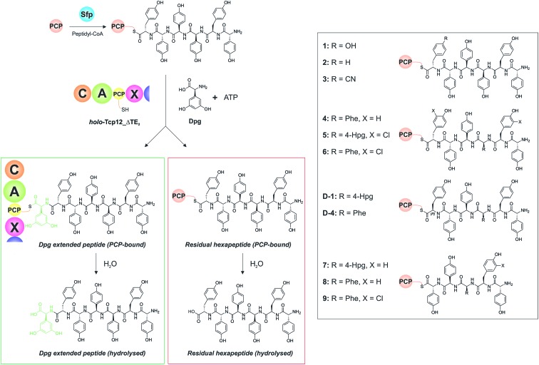
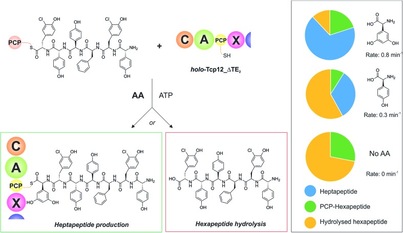
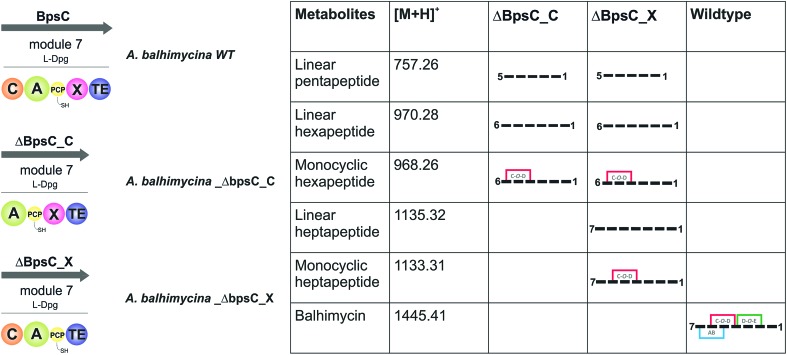
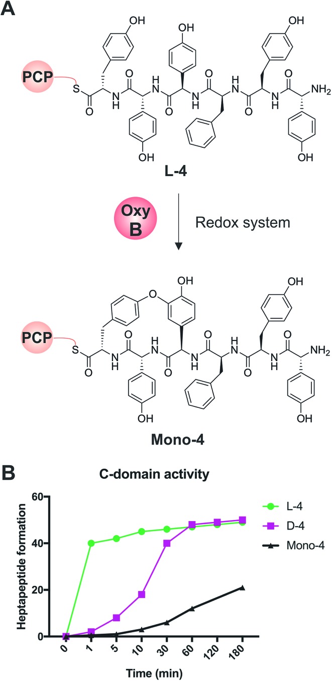
Non-ribosomal peptide synthesis is a highly important biosynthetic pathway for the formation of many secondary metabolites of medical relevance. Due to the challenges associated with the chemical synthesis of many of the products of these assembly lines, understanding the activity and selectivity of non-ribosomal peptide synthetase (NRPS) machineries is an essential step towards the redesign of such machineries to produce new bioactive peptides. Whilst the selectivity of the adenylation domains responsible for amino acid activation during NRPS synthesis has been widely studied, the selectivity of the essential peptide bond forming domains - known as condensation domains - is not well understood. Here, we present the results of a combination of in vitro and in vivo investigations into the final condensation domain from the NRPS machinery that produces the glycopeptide antibiotics (GPAs). Our results show that this condensation domain is tolerant for a range of peptide substrates and even those with unnatural stereochemistry of the peptide C-terminus, which is in contrast to the widely ascribed role of these domains as a stereochemical gatekeeper during NRPS synthesis. Furthermore, we show that this condensation domain has a significant preference for linear peptide substrates over crosslinked peptides, which indicates that the GPA crosslinking cascade targets the heptapeptide bound to the final module of the NRPS machinery and reinforces the role of the unique GPA X-domain in this process. Finally, we demonstrate that the peptide bond forming activity of this condensation domain is coupled to the rate of amino acid activation performed by the subsequent adenylation domain. This is a significant result with implications for NRPS redesign, as it indicates that the rate of amino acid activation of modified adenylation domains must be maintained to prevent unwanted peptide hydrolysis from the NRPS due to a loss of the productive coupling of amino acid selection and peptide bond formation. Taken together, our results indicate that assessing condensation domain activity is a vital step in not only understanding the biosynthetic logic and timing of NRPS-mediated peptide assembly, but also the rules which redesign efforts must obey in order to successfully produce functional, modified NRPS assembly lines.


Figures

Fig. 1
Fig. 1. Schematic representation of peptide bond formation as performed by a NRPS extension module containing an epimerisation domain. Amino acid selection and subsequent activation are performed by the adenylation (A)-domain (first panel, starting from the left), after which the amino acid is transferred onto the PPant moiety of the neighbouring carrier protein (CP) domain. Following this, the aminoacyl-CP then acts as the acceptor in peptide bond formation performed by the upstream condensation domain (second panel). At this point, the peptide present on the upstream donor CP is transferred onto the acceptor aminoacyl-CP, extending the peptide by one residue. Depending on the stereochemistry required at the C-terminal position of the peptide, an epimerisation domain can alter the standard l-configuration into the non-natural d-form (third panel). The stereochemical state of the peptide is then assessed by the downstream C-domain, where the peptidyl-CP now acts as the donor substrate for the next round of peptide bond formation (fourth panel).
Fig. 2
Fig. 2. Biosynthetic scheme for the glycopeptide antibiotics (GPAs), exemplified for teicoplanin (type-IV GPA, upper panel) as well as related GPA structures relevant for this work actinoidin (type-II GPA, lower left) and balhimycin (type-I GPA lower right). Type-III GPAs possess the same core peptide sequence as type-IV GPAs. In GPA biosynthesis, the NRPS-mediated synthesis of a linear heptapeptide precursor is followed by an oxidative peptide cyclisation cascade of cytochrome P450 (Oxy) enzymes, which transform the linear peptide into its rigid, active form whilst the peptide remains bound to the NRPS machinery. In the biosynthesis of the three GPAs indicated here, the NRPS machinery remains the same from a domain and module perspective: the main differences between these GPA biosynthetic machineries are the number of Oxy enzymes and hence crosslinks installed in the cyclic peptide (3 – balhimycin/actinoidin; or 4 – teicoplanin), the presence of 3 (balhimycin) or 4 (actinoidin/teicoplanin) NRPS-encoding proteins, and the residues contained within the peptide that are dictated by the selectivity of the A-domains.
Fig. 3
Fig. 3. Construction of the mutant A. balhimycina_ΔbpsCX. Schematic representation of the deletion of the X domain. Domain arrangement shown for the balhimycin NRPS. NRPS domain descriptions: C, condensation; A, adenylation; PCP, peptidyl carrier protein; E, epimerisation; X, P450 (Oxy) recruitment; TE, thioesterase. ermE, erythromycin resistance gene. bpsCXleft (2079 bp): red; bpsCXright (1916): green. To obtain the deletion mutant A. balhimycina_ΔbpsCX a double crossover via homologous recombination is required.
Fig. 4
Fig. 4. Characterisation of the amino acid selection and activation characteristics of the final NRPS module from teicoplanin biosynthesis (Tcp12). (A) Alternate constructs of Tcp12, the final module from the teicoplanin NRPS, were designed in order to remove the C-terminal TE-domain in order to prevent unwanted peptide hydrolysis during subsequent C-domain assays (Tcp12_ΔTE1, Tcp12_ΔTE2, Tcp12_ΔTE3). NRPS domain descriptions: C, condensation; A, adenylation; PCP, peptidyl carrier protein; X, P450 (Oxy) recruitment; TE, thioesterase. (B) Amino acid selectivity of the A-domain of the apo-Tcp12 protein for the natural substrate Dpg (l-3,5-dihydroxyphenylglycine) as well as related Phg substrates (4-Hpg: l-4-hydroxylphenylglycine; 3-Hpg: l-3-hydroxylphenylglycine; 4-Hpg: l-phenylglycine).
Fig. 5
Fig. 5. Condensation domain assay for the final module of the teicoplanin NRPS. Initially, peptidyl-CoA substrates prepared by solid phase peptide synthesis are loaded onto the isolated PCP domain from the preceding module using the promiscuous phosphopantetheinyl transferase Sfp (top left), after which these substrates are then added to the Tcp12_ΔTE2 construct along with Dpg and ATP in order to assess the formation of Dpg-extended peptide products. Products peptides are extended by the addition of Dpg through the actions of Tcp12_ΔTE2 (green box, residual starting peptide shown in the red box). All peptides can either remain PCP-bound at the end of the assay (where they are then analysed as their methylamides through the addition of methylamine) or they can be hydrolysed from the PCP. Peptide structures synthesised and used as substrates in these assays are shown in the boxed area on the right of the figure (1–9, D-1, D-4). NRPS domain descriptions: C, condensation; A, adenylation; PCP, peptidyl carrier protein; X, P450 (Oxy) recruitment.
Fig. 6
Fig. 6. Determining the effect of downstream A-domain rate on upstream C-domain peptide bond formation by varying the amino acid provided during the peptide bond forming step. Summary of the assay (left), in which peptide 4 was loaded onto PCP6 and used to reconstitute heptapeptide bond formation in the presence of the natural A-domain amino acid substrate Dpg, a substrate activated ∼2.5× more slowly (4-Hpg) and no amino acid substrate. Results (box on right) show that an increase in hydrolysed hexapeptide starting material (shown in yellow; residual PCP-bound hexapeptide shown in green) correlates to the presence of a poorer (or no) suitable A-domain substrate, with a decrease in extended peptide product (shown in blue).
Fig. 7
Fig. 7. Analysis of the roles of the C- and X-domains within the final NRPS module from balhimycin biosynthesis as assessed through the isolation and analysis of the peptide products formed by the resultant mutant producer strains in which these domains had been removed. Results show that the initial cyclisation step performed by OxyB can occur on the peptide at the hexapeptide stage, which raises the question of cyclisation vs. heptapeptide formation during GPA biosynthesis. NRPS domain descriptions: C, condensation; A, adenylation; PCP, peptidyl carrier protein; X, P450 (Oxy) recruitment; TE, thioesterase.
Fig. 8
Fig. 8. Analysis of the acceptance of the Tcp12 C-domain for different hexapeptide substrates, either the standard linear hexapeptide (L-4), a peptide bearing altered stereochemistry at the C-terminal residue (D-4) and a monocyclic version of L-4 (Mono-4) formed through the actions of OxyBvan. (A) Initial formation of Mono-4 was performed on PCP6 using OxyBvan and an appropriate redox system. (B) Comparison of the rate of cyclisation of these three peptides under identical reaction conditions shows that the L-4 peptide is accepted significantly faster than the d-configured form of this peptide (half conversion at 30 seconds vs. 15 minutes), whilst the cyclised peptide Mono-4 is only accepted for peptide extension at a very slow rate (half conversion > 200 minutes).

References

    1. Weissman K. J. Nat. Prod. Rep. 2016;33:203–230. - PubMed
    1. Klaus M., Grininger M. Nat. Prod. Rep. 2018 doi: 10.1039/c8np00030a. - DOI - PubMed
    1. Süssmuth R. D., Mainz A. Angew. Chem., Int. Ed. 2017;56:3770–3821. - PubMed
    1. Winn M., Fyans J. K., Zhuo Y., Micklefield J. Nat. Prod. Rep. 2016;33:317–347. - PubMed
    1. Payne J. A. E., Schoppet M., Hansen M. H., Cryle M. J. Mol. BioSyst. 2017;13:9–22. - PubMed