Review
. 2019 Apr;8(8):e1801578.
doi: 10.1002/adhm.201801578. Epub 2019 Feb 4.

Extracellular Matrix-Based Strategies for Immunomodulatory Biomaterials Engineering

Affiliations
Review

Extracellular Matrix-Based Strategies for Immunomodulatory Biomaterials Engineering

Andrew T Rowley et al. Adv Healthc Mater. 2019 Apr.

Abstract

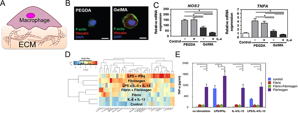
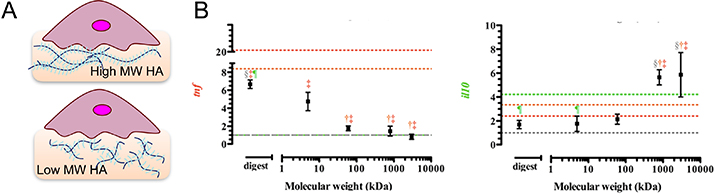
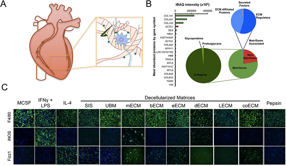
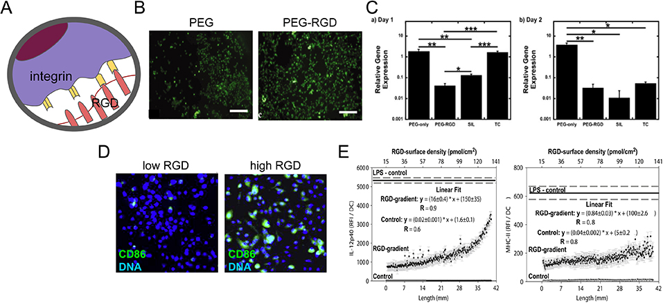
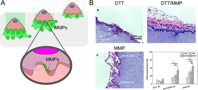
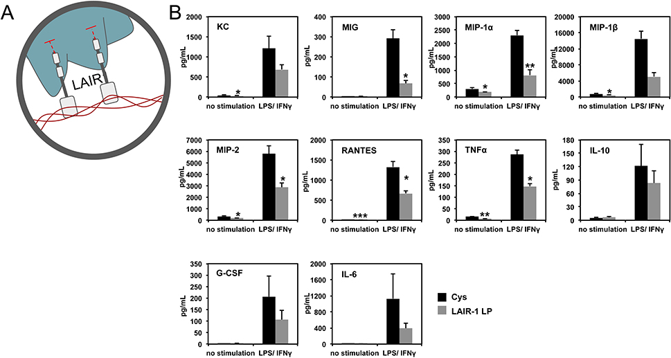
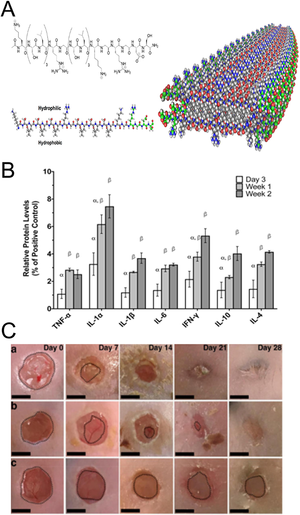
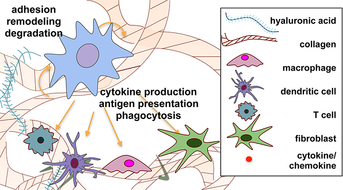
The extracellular matrix (ECM) is a complex and dynamic structural scaffold for cells within tissues and plays an important role in regulating cell function. Recently it has become appreciated that the ECM contains bioactive motifs that can directly modulate immune responses. This review describes strategies for engineering immunomodulatory biomaterials that utilize natural ECM-derived molecules and have the potential to harness the immune system for applications ranging from tissue regeneration to drug delivery. A top-down approach utilizes full-length ECM proteins, including collagen, fibrin, or hyaluronic acid-based materials, as well as matrices derived from decellularized tissue. These materials have the benefit of maintaining natural conformation and structure but are often heterogeneous and encumber precise control. By contrast, a bottom-up approach leverages immunomodulatory domains, such as Arg-Gly-Asp (RGD), matrix metalloproteinase (MMP)-sensitive peptides, or leukocyte-associated immunoglobulin-like receptor-1(LAIR-1) ligands, by incorporating them into synthetic materials. These materials have tunable control over immune cell functions and allow for combinatorial approaches. However, the synthetic approach lacks the full natural context of the original ECM protein. These two approaches provide a broad range of engineering techniques for immunomodulation through material interactions and hold the potential for the development of future therapeutic applications.

Keywords: biomaterials; cytokines; extracellular matrix; immunomodulation; inflammation; tissue engineering; wound repair.


Conflict of interest statement

Conflicts of Interest

The authors have no conflicts of interest to declare.

Figures

Figure 1.
Figure 1.
Schematic representation of ECM-immune cell interactions. Interactions include LAIR1-collagen interaction that inhibit inflammatory signaling, MMPs that drive matrix degradation at cleavage motifs, and RGD that facilitates cellular adhesion to ECM via integrin binding.
Figure 2.
Figure 2.
Collagen- and fibrin-based materials modulate the macrophage inflammatory response. (A) Schematic of immune cell interaction with ECM-based materials. (B) Confocal images of THP-1 macrophage expression of adhesion molecule vinculin on nonadhesive polyethylene glycol diacrylate hydrogel (PEGDA, left) and adhesive gelatin hydrogel (GelMA, right). Scale bar is 10 μm. (C) mRNA expression levels of inflammatory markers NOS2 and TNFA in macrophages cultured on PEGDA vs. GelMA with or without IL-4 treatment. (B) and (C) are adapted from Cha et al., 2017.[22] (D) Z-score heatmap of inflammatory cytokines secreted by macrophages cultured on fibrin or polystyrene and stimulated with LPS, IFNγ, IL-4, IL-13, and/or fibrinogen. Red indicates relatively high levels of secretion and blue indicates low levels of secretion. (E) TNFα secretion by BMDM cultured on control polystyrene (blue and purple), fibrin (red and green), and/or fibrinogen (green and purple) in the indicated stimulation conditions. (D) and (E) are adapted from Hsieh et al. 2017.[37]
Figure 3.
Figure 3.
Effect of HA molecular weight on macrophage polarization. (A) Schematic of immune cell cultured on HA hydrogels of varied molecular weights. (B) Gene expression of Il10 and Tnfα in mouse sarcoma macrophages J774A.1 stimulated with IL-4 and HA of different molecular weights for 24 h. Adapted from Rayahin et al., 2015.[53]
Figure 4.
Figure 4.
Tissue source affects composition and immunomodulatory effects of decellularized matrices. (A) Schematic of extraction of decellularized matrix. (B) Composition of commercial porcine urinary bladder matrix (MicroMatrix™) as determined by mass spectrometry. Adapted from Sadtler et al., 2017.[71] (C) Immunofluorescence images of F4/80 (pan macrophage), iNOS (M1 marker) and Fizz1 (M2 marker) in macrophages cultured on polystyrene with cytokines or pepsin, or solubilized decellularized ECM derived from various tissues: SIS (small intestinal submucosa), UBM (urinary bladder matrix), mECM (skeletal muscle ECM), bECM (brain ECM), eECM (esophageal ECM), dECM (dermal ECM), lECM (liver ECM), coECM (colonic ECM). Adapted from Dziki et al., 2017.[78]
Figure 5.
Figure 5.
RGD facilitates cellular adhesion and differentially modulates the activation of myeloid cells. (A) Schematic representation of cellular adhesion facilitated by RGD domains in the ECM and cellular integrins. (B) Immunofluorescence images of calcein AM-stained BMDMs on PEG only or RGD-PEG surfaces. Scale bar is 200 μm. (C) Relative gene expression of Tnfa in BMDMs at days one (left) and two (right), seeded onto PEG-only, PEG-RGD, medical grade silicon (SIL) and tissue culture polystyrene (TC) surfaces. Gene expression was normalized to housekeeping gene L32. (B) and (C) are adapted from Lynn et al., 2010.[88] (D) Immunofluorescence image of CD86 immunostained (green) and dendritic cell nuclei stained with DAPI (blue) DCs on low and RGD densities. (E) Expression of DC production of IL-12p40 and stimulatory molecule MHC-II when cultured for 24 h a gradient hydrogel with indicated RGD peptide surface density. The solid line represents the background-corrected relative fluorescence intensity of DCs cultured in the presence of LPS with the dashed line indicating the standard error. Data sets were linear curve fit and equations with the obtained parameters are shown. (D) and (E) are adapted from Acharya et al., 2010.[89]
Figure 6.
Figure 6.
MMP sensitive peptides facilitate immune-mediated matrix degradation. (A) Schematic representation of immune cell infiltration facilitated by matrix degradation via MMPs. (B) H&E stained sections of dithiothreitol (DTT) crosslinked hydrogels with increasing the MMP sensitive peptide crosslinker content (from 0 to 100%) implanted subcutaneously in mice for a total of 21 days and quantification (bottom right) of cell invasion distance in the different materials at 3, 6, 12, and 21 days. Adapted from Yu et al., 2018.[131]
Figure 7.
Figure 7.
LAIR1 ligands in collagen modulate inflammatory cytokine production. (A) Schematic representation of LAIR-1 binding to collagen. (B) Levels of secreted cytokines from BMDM cultured on cysteine (control) or LAIR-1 ligand peptide (LP) coated surfaces, with or without LPS/IFNγ stimulation. Adapted from Kim et al., 2017.[120]
Figure 8.
Figure 8.
MDPs are self-assembled molecules that allow for the incorporation of multiple ECM derived peptides. (A) Schematic depiction of self assembly of A-B-A block motif peptides into the basic fiber building block of MDPs, adapted from Kumar et al, 2015.[140] B) Relative levels of pro- and anti-inflammatory cytokines present in dorsal tissues after MDP subcutaneous injection into rats at different time points.[139] (C) Gross morphometry images of skin wounds were treated with MDP hydrogel (top), IntraSite (middle), and buffer control (bottom). Scale bars are 5mm Adapted from Carrejo et al., 2018.[126]
Figure 9.
Figure 9.
Schematic representation of dynamic cell-matrix crosstalk during immune cell-matrix interactions. Material-cell crosstalk is represented through degradation of matrix by enzymes produced by cells, the production of ECM proteins by cells, and the immunomodulatory domains in the ECM described in this review. Cellular crosstalk is depicted via the production of, and interaction with, cytokines and chemokines by the various cell types.

References

    1. Vaday GG, Lider O. Extracellular matrix moieties, cytokines, and enzymes: dynamic effects on immune cell behavior and inflammation. J Leukoc Biol. 2000;67(2):149–159. - PubMed
    1. Singh A, Peppas NA. Hydrogels and Scaffolds for Immunomodulation. Advanced Materials. 2014;26(38):6530–6541. - PMC - PubMed
    1. Dziki JL, Huleihel L, Scarritt ME, Badylak SF. Extracellular Matrix Bioscaffolds as Immunomodulatory Biomaterials. Tissue Engineering Part A. 2017;23(19–20):1152–1159. - PMC - PubMed
    1. Midwood KS, Williams LV, Schwarzbauer JE. Tissue repair and the dynamics of the extracellular matrix. The International Journal of Biochemistry & Cell Biology. 2004;36(6):1031–1037. - PubMed
    1. Taraballi F, Sushnitha M, Tsao C, et al. Biomimetic Tissue Engineering: Tuning the Immune and Inflammatory Response to Implantable Biomaterials. Advanced Healthcare Materials. 2018;7(17):1800490. - PubMed

Publication types

Substances