Review
. 2019 Mar;8(5):e1801217.
doi: 10.1002/adhm.201801217. Epub 2019 Feb 4.

Decellularized Extracellular Matrix Materials for Cardiac Repair and Regeneration

Affiliations
Review

Decellularized Extracellular Matrix Materials for Cardiac Repair and Regeneration

Donald Bejleri et al. Adv Healthc Mater. 2019 Mar.

Abstract

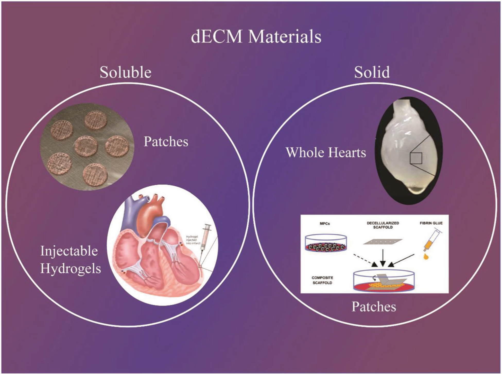
Decellularized extracellular matrix (dECM) is a promising biomaterial for repairing cardiovascular tissue, as dECM most effectively captures the complex array of proteins, glycosaminoglycans, proteoglycans, and many other matrix components that are found in native tissue, providing ideal cues for regeneration and repair of damaged myocardium. dECM can be used in a variety of forms, such as solid scaffolds that maintain native matrix structure, or as soluble materials that can form injectable hydrogels for tissue repair. dECM has found recent success in many regeneration and repair therapies, such as for musculoskeletal, neural, and liver tissues. This review focuses on dECM in the context of cardiovascular applications, with variations in tissue and species sourcing, and specifically discusses advances in solid and soluble dECM development, in vitro studies, in vivo implementation, and clinical translation.

Keywords: cardiac patches; decellularized; extracellular matrices; injectable hydrogels.


Conflict of interest statement

Conflict of Interest

The authors declare no conflict of interest.

Figures

Figure 1.
Figure 1.
Main terminology used to differentiation dECM technologies. Examples: (Top left - patches) Reproduced with permission.[103] Copyright 2008, Springer Nature. (Bottom left - injectable hydrogels) Reproduced with permission.[131] Copyright 2011, National Academy of Sciences. (Top right - whole hearts) Reproduced with permission.[37] Copyright 2018, Wiley-VCH. (Bottom right - patches) Reproduced with permission.[73] Copyright 2016, Elsevier.
Figure 2.
Figure 2.
Biochemical and structural characterization of fetal (E18) and adult bioscaffolds. A) Side-by-side comparison of immunofluorescence for main ECM components before and after decellularization. Scale bar, 100 μm. B) Quantification of fibronectin and mature collagen type I by Western blot. C,D) Detail of the basement membrane and pericellular matrix obtained following C) whole mount immunostaining and optical clearing and by D) TEM. Scale bar: C) 50 μm and D) 2 μm. E) Representative images of fetal (E18) and adult decellularized scaffolds surface by SEM. Scale bars are identified in the image. # nanofibrils (<0.1 μm), *thick microfibers (>0.3 μm). F) Bar graph shows the percentage of fibrils with different diameters following quantification on SEM images. G) Representative images of collagen fibers obtained by SEM (left) and TEM (right) on tissue ultrathin sections. Bar graph shows the mean diameter of fibril units. Scale bar: 1 μm. Representative images and quantitative data were obtained from two or three (Western blot) independent experiments. Data are expressed as mean ± SEM. Student’s t-test, two-tailed, *p < 0.05, **p < 0.01, ***p < 0.001. Reproduced with permission.[14] Copyright 2016, Elsevier.
Figure 3.
Figure 3.
a–c) Photographs of cadaveric rat hearts mounted on a Langendorff apparatus. Ao, aorta; LA, left atrium; LV, left ventricle; RA, right atrium; RV, right ventricle. Retrograde perfusion of cadaveric rat heart using A) PEG, B) Triton-X-100 or C) SDS over 12 h. The heart becomes more translucent as cellular material is washed out from the right ventricle, then the atria and finally the left ventricle. d,e) Corresponding H&E staining of thin sections from LV of rat hearts perfused with D) PEG or E) Triton-X-100, showing incomplete decellularization. Hearts treated with PEG or Triton-X-100 retained nuclei and myofibers. Scale bars: 200 μm. F) H&E staining of thin section of SDS-treated heart showing no intact cells or nuclei. Scale bar: 200 μm. All three protocols maintain large vasculature conduits (black asterisks). G) Immunofluorescent staining of cadaveric and SDS-decellularized rat heart thin sections showing the presence or absence of DAPI-positive nuclei (purple), cardiac α-myosin heavy chain (green) or sarcomeric α-actin (red). Nuclei and contractile proteins were not detected in decellularized constructs. Scale bars: 50 μm. Reproduced with permission.[37] Copyright 2008, Springer Nature.
Figure 4.
Figure 4.
Cell delivery platform. A) Composite scaffolds assembled from thin sheets of decellularized human myocardium and fibrin hydrogel were seeded with MPCs, cultured in vitro with or without TGF-β conditioning, and implanted into nude rat models of acute and chronic ischemia. After 4 week, heart function was evaluated by echocardiography ex vivo analyses. B) The three axes of the heart (r, q, and z), the directions for sectioning (apex to base, circumferential, and radial), and the short axis of myocytes seen in circumferential sections. C–F) Scanning electron micrographs of decellularized scaffolds: apex to base sections (C; 796×), radial sections (D; 986×), and circumferential sections (E; 1000×; F, 2500 ×). Staining of extracellular matrix components circumferential sections of native and decellularized tissue: G) collagen IV, H) laminin, I) vitronectin, and J) fibronectin. (Scale bar: 250 μm.) K) Average pore diameters in decellularized scaffolds. L) Similar properties of native (circumferential, blue; radial, pink) and decellularized (circumferential, green; radial, purple) tissue measured in uniaxial tensile tests (tensile moduli calculated at 20% strain). Reproduced with permission.[73] Copyright 2011, National Academy of Sciences.
Figure 5.
Figure 5.
Printing overview. A) Bioink preparation involved combining cECM, hCPCs, and GelMA to form naturally derived and cell-laden materials for printing. B) Printing methodology involved cooling the bioink to 10 °C in the 3D bioprinter barrels to allow GelMA polymerization for improved printability. Patches were printed with infill patterns of 90° intersecting filaments and contour. Patches were polymerized via white light to induce radical polymerization of GelMA, followed by incubation at 37 °C for at least 1 h to induce cECM polymerization. C) Patch implementation will involve pericardially inserting the patch to the RV of pediatric patients, where the patch will release key regenerative paracrine factors. Reproduced with permission.[103] Copyright 2018, Wiley-VCH.
Figure 6.
Figure 6.
Schematic of prevascularized stem cell patch. A) Illustration of 3D cell printing system, and B) macroscopic view of the printer. C) Illustration of prevascularized stem cell patch including multiple cell-laden bioinks and supporting PCL polymer. D) Fabricated patch including the two types of cell-laden bioink and PCL supporting layer [Scale bar (top left), 1 mm; Scale bar (bottom), 200 μm]. All bioinks were composed of cECM. Reproduced with permission.[123] Copyright 2017, Elsevier.
Figure 7.
Figure 7.
Effects of myocardial matrix hydrogel after myocardial infarction: mechanisms underlying the functional benefits. Injection of myocardial matrix 1 week after myocardial infarction (MI) into the infarcted area induced various tissue level changes that reduced negative left ventricular remodeling and improved hemodynamics. Altering these key pathways created a pro-regenerative environment, potentially preventing or slowing development of heart failure. Reproduced with permission.[132] Copyright 2016, Elsevier.
Figure 8.
Figure 8.
Histological and immunohistochemical analysis after in vivo scaffold implantation. A) Representative images (top to bottom) of H/E, Movat’s pentachrome, Movat’s pentachrome for simultaneous collagen and mucopolysaccharide acid staining and Gallego’s modified trichrome. Scale bar = 100 μm. B) Movat’s pentachrome of the scaffolds from the Per-ATMSCs, Myo-ATMSCs, Per-MI, and Myo-MI groups displaying the presence of arterial blood vessels (red arrows), veins (blue arrows), and nerve fibers (yellow arrows). Scale bar = 50 μm. C) Representative SEM images of scaffolds from Per-MI and Myo-MI experimental groups after sacrifice. D) Immunohistochemical images of the scaffolds against IsoB4 (green), SMA (red), and elastin (white) antibodies confirming the presence of arteriolar blood vessels positive for SMA. Nuclei appear counterstained in blue. Scale bars = 50 μm. Abbreviations: Per-MI (cell-free pericardial scaffold), Myo-MI (cell-free myocardial scaffold), Per-ATMSCs (adipose tissue mesenchymal stem cell-enriched pericardial scaffold), Myo-ATMSCs (adipose tissue mesenchymal stem cell-enriched myocardial scaffold). Reproduced with permission.[42] Copyright 2018, Springer Nature.

References

    1. Radisic M, Christman KL, Mayo Clin. Proc 2013, 88, 884. - PMC - PubMed
    1. Taylor DA, Sampaio LC, Ferdous Z, Gobin AS, Taite LJ, Acta Biomater. 2018, 74, 74. - PubMed
    1. Spang MT, Christman KL, Acta Biomater. 2018, 68, 1. - PMC - PubMed
    1. Garreta E, Oria R, Tarantino C, Pla-Roca M, Prado P, Fernandez-Aviles F, Campistol JM, Samitier J, Montserrat N, Mater. Today 2017, 20, 166.
    1. Landa N, Miller L, Feinberg MS, Holbova R, Shachar M, Freeman I, Cohen S, Leor J, Circulation 2008, 117, 1388. - PubMed

Publication types

MeSH terms

Substances