Review
. 2019 Feb 18;48(4):971-988.
doi: 10.1039/c8cs00157j.

Molecular methods for assessment of non-covalent metallodrug-DNA interactions

Affiliations
Review

Molecular methods for assessment of non-covalent metallodrug-DNA interactions

Andrew Kellett et al. Chem Soc Rev. .

Abstract

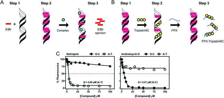
The binding of small molecule metallodrugs to discrete regions of nucleic acids is an important branch of medicinal chemistry and the nature of these interactions, allied with sequence selectivity, forms part of the backbone of modern medicinal inorganic chemistry research. In this tutorial review we describe a range of molecular methods currently employed within our laboratories to explore novel metallodrug-DNA interactions. At the outset, an introduction to DNA from a structural perspective is provided along with descriptions of non-covalent DNA recognition focusing on intercalation, insertion, and phosphate binding. Molecular methods, described from a non-expert perspective, to identify non-covalent and pre-associative nucleic acid recognition are then demonstrated using a variety of techniques including direct (non-optical) and indirect (optical) methods. Direct methods include: X-ray crystallography; NMR spectroscopy; mass spectrometry; and viscosity while indirect approaches detail: competitive inhibition experiments; fluorescence and absorbance spectroscopy; circular dichroism; and electrophoresis-based techniques. For each method described we provide an overview of the technique, a detailed examination of results obtained and relevant follow-on of advanced biophysical/analytical techniques. To achieve this, a selection of relevant copper(ii) and platinum(ii) complexes developed within our laboratories are discussed and are compared, where possible, to classical DNA binding agents. Applying these molecular methods enables us to determine structure-activity factors important to rational metallodrug design. In many cases, combinations of molecular methods are required to comprehensively elucidate new metallodrug-DNA interactions and, from a drug discovery perspective, coupling this data with cellular responses helps to inform understanding of how metallodrug-DNA binding interactions manifest cytotoxic action.


Figures

Fig. 1
Fig. 1. (A) X-ray structures of A-, B- and Z-DNA from PDB files 1VJ4, ; 1BNA and ; 2DCG respectively, (B) hydrogen bonding between C–G and T–A base pairs, (C) summary of structural differences between A-, B- and Z-DNA; (D) conformational preferences of the 2′-deoxyribose rings of DNA taken from ; 1BNA and ; 2DGC with respective anti- and syn-conformations of guanine nucleobase and (E) top down view of stacking interactions in TA and AT steps from PDB ; 167D (d(CCATTAATGG)2). Hydrogen bonds are shown as dashed yellow lines; the angle between the vectors representing the two N3(T) → N1(A) hydrogen bonds is 118° for the TA step and 160° for the AT step.
Fig. 2
Fig. 2. (A) Cu-Phen and ternary copper(ii) phenanthrene complexes Cu-DPQ-Phen, Cu-DPPZ-Phen and Cu-DPPN-Phen; (B) di-nuclear copper(ii) terephthalate (Cu-Terph) and octanedioate (Cu-Oda) complexes; (C) square planar copper(ii) complex Cu-Ph-Phen incorporating o-phthalate and 1,10-phenanthroline; (D) polynuclear platinum complexes (PPCs) [{transPtCl(NH3)2}2-μ-{trans-Pt(NH3)2(NH2(CH2)6NH2)2}]4+ (Triplatin, BBR3464), [{Pt(NH3)3}2-μ-{trans-Pt(NH3)2(NH2(CH2)6NH2)2}]6+ (AH44), and [{transPt(NH3)2(NH2(CH2)6NH3)}2-μ-({trans-Pt(NH3)2(NH2(CH2)6NH2)2})]8+ (TriplatinNC); and (E) Minor groove binding agents 4′,6-diamidino-2-phenylindole (DAPI), netropsin (Net), Hoechst 33258; (F) major groove binding methyl green (MG); and (G). planar heterocyclic intercalators propidium iodide (PI), ethidium bromide (EtBr) and doxorubicin (Dox).
Fig. 3
Fig. 3. Molecular structures of selected Pt(ii), Rh(iii) and Ru(ii) intercalating and insertion complexes.
Fig. 4
Fig. 4. Intercalation and insertion. (A) Δ-α-[Rh{(R,R)-Me2trien}phi]3+ intercalated into 5′-G(5|U)TGCAAC-3′ with additional stabilisation by H-bonds from the ancillary ligand (PDB ; 454d), black lines indicate hydrogen bonds and (B) Δ-[Rh(bpy)2(chrysi)]3+ inserted into (5′-CGGAAATTCCCG-3′), displacing a mismatched AC pair shown in green (PDB ; 2O1I) b; (C) intercalation of Λ-[Ru(phen)2(dppz)]2+ into d(CCGGTACCGG)2 (PDB ; 3U38); (D) Λ-[Ru(TAP)2(dppz)]2+ bound to d(CCGGATCCGG)2 by semi-intercalation (PDB ; 4YMC); TriplatinNC bound to Dickerson–Drew dodecamer (B-DNA) through backbone tracking (E) and groove spanning (F) interactions. (N–H···OP hydrogen bonds shown as dashed red lines); and (G) three-way DNA junction recognition by a metallosupramolecular helicate (Fe2L3)4+ where L = C25H20N4 (PDB ; 2ET0).
Fig. 5
Fig. 5. {1H,15N} HSQC NMR of TriplatinNC (left) and Dickerson–Drew Duplex (DDD, right). Satellites from 1J(15N–195Pt) are clearly visible. Adapted with permission from Qu et al. Reproduced from ref. 32 with permission from the Royal Society of Chemistry, copyright 2015.
Fig. 6
Fig. 6. ESI-MS/MS schematic of free (top) and PPC (either TriplatinNC or AH44) adducted (bottom) 5′-d(TCTCCCAGCGTGCGCCAT) at 100 and 120 V of collisional energy, respectively. Fragmentation of the glycosidic bonds is prevalent throughout the free, with the region of enhanced stability in teal. The associated fragment ions (w82–, w92–, and a9–a12 using standard McLuckey nomenclature) are absent in the adduct indicating the area of PPC binding.
Fig. 7
Fig. 7. Relative viscosity values of organic and inorganic compounds bound to duplex stDNA.
Fig. 8
Fig. 8. (A) CD profile of B-form salmon testes DNA (stDNA) highlighting wavelengths of interest including 210, 220, 246 and 268 nm; (B) change in ellipticity of stDNA at 220 nm in the presence increasing ratios of methyl green (MG) and di-nuclear Cu2TPNap; (C) B → Z NaCl titration of alternating co-polymer poly[d(G–C)2]; and (D) CD spectra of 12mer sequences d(GCCGGTACCGGC)2, d(GCCGGATCCGGC)2 and overlay of d(GCTTTATAAAGC)2 and d(GCUUUAUAAAGC)2 sequences in the presence of Cu-Oda.
Fig. 9
Fig. 9. (A) Indirect fluorescence displacement assay using ethidium bromide as a reporter molecule to determine apparent binding constants (Kapp) of non-fluorescent DNA-binding agents; (B) FPX-TriPtNC binding assessment assay; and (C) fluorescence quenching of limited bound intercalator (EtBr) to poly[d(G–C)2] and poly[d(A–T)2] upon titration of netropsin and actinomycin D. Reproduced from ref. 37 with permission from the American Chemical Society, copyright 2014.
Fig. 10
Fig. 10. (A) Cartoon illustration of superhelical (SC), open circular (OC) and linear (L) DNA and agarose gel electrophoresis representation of respective DNA forms; (B) topoisomerase I mediated DNA relaxation assay in the presence of intercalating agent EtBr; (C) table of selected free radical scavengersa and intracellular antioxidantsb; and (D) gel profile of plasmid DNA treated with Cu-Ph-Phen in the absence (lanes 2–5) and presence of radical-specific antioxidants and trapping agents (lanes 6–21). Reproduced from ref. 50 with permission from the American Chemical Society, copyright 2016.
Fig. 11
Fig. 11. (A) Double stranded DNA “on-chip” protocol developed within our research laboratories used to assess (i) oxidative DNA damage and (ii) PPC non-covalent binding interactions using the Agilent BioAnalyser 2100 platform; (B) melphalan alkylation assay using piperidine and heat to induce thermolabbile scission and (C) minor groove protection assay polynuclear platinum complexes (PPC).
None
From left to right: Dr Zara Molphy, Dr Andrew Kellett, Prof. Nicholas P. Farrell, Dr Creina Slator, and Prof. Vickie McKee

References

    1. Barry N. P. E., Sadler P. J. Chem. Commun. 2013;49:5106–5131. - PubMed
    1. Chargaff E. Science. 1971;172:637–642. - PubMed
    1. Wing R., Drew H., Takano T., Broka C., Tanaka S., Itakura K., Dickerson R. E. Nature. 1980;287:755–758. - PubMed
    1. Rich A., Zhang S. Nat. Rev. Genet. 2003;4:566–572. - PubMed
    1. Drew H. R., Dickerson R. E. J. Mol. Biol. 1981;152:723–736. - PubMed