. 2019 Feb 26;19(5):778-788.
doi: 10.1039/c8lc01200h.

Modular soft robotic microdevices for dexterous biomanipulation

Affiliations

Modular soft robotic microdevices for dexterous biomanipulation

Berna Özkale et al. Lab Chip. .

Abstract

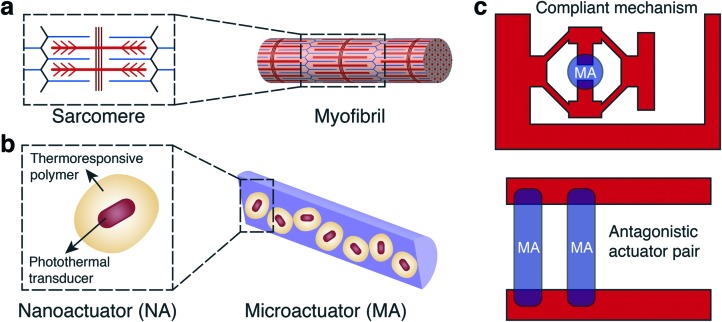
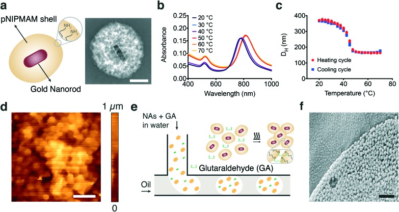
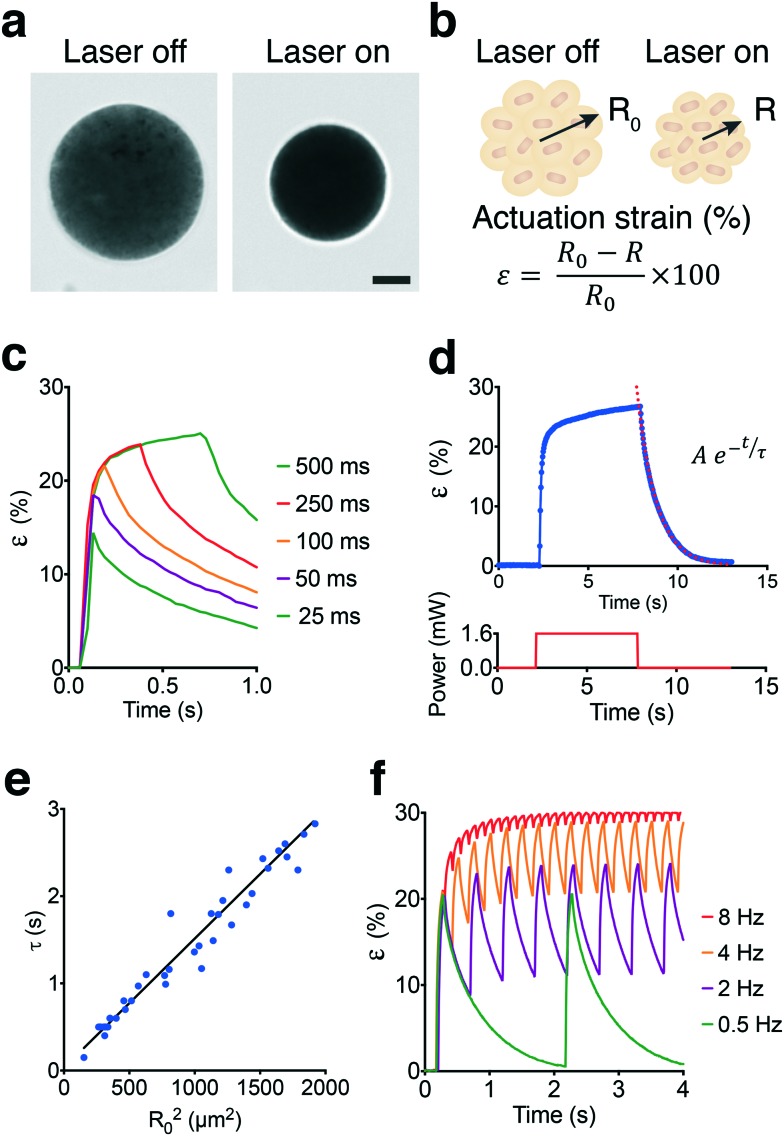
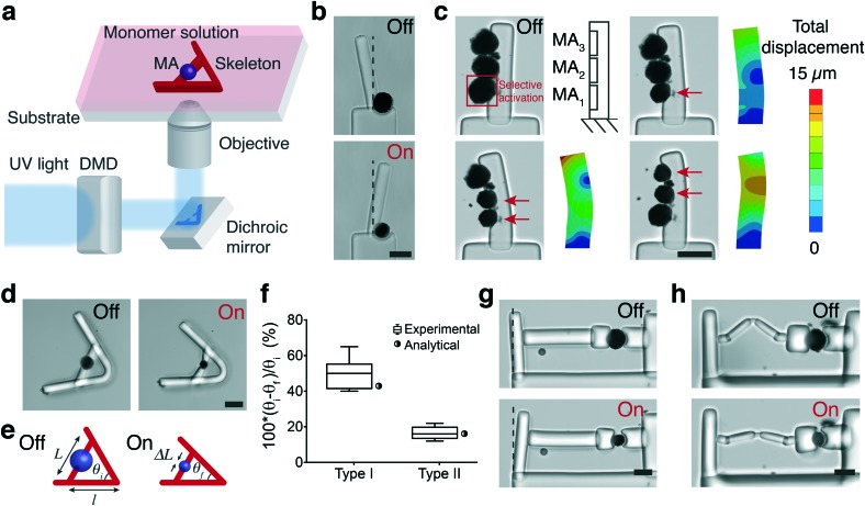
We present a methodology for building biologically inspired, soft microelectromechanical systems (MEMS) devices. Our strategy combines several advanced techniques including programmable colloidal self-assembly, light-harvesting with plasmonic nanotransducers, and in situ polymerization of compliant hydrogel mechanisms. We synthesize optomechanical microactuators using a template-assisted microfluidic approach in which gold nanorods coated with thermoresponsive poly(N-isopropylmethacrylamide) (pNIPMAM) polymer function as nanoscale building blocks. The resulting microactuators exhibit mechanical properties (4.8 ± 2.1 kPa stiffness) and performance metrics (relative stroke up to 0.3 and stress up to 10 kPa) that are comparable to that of bioengineered muscular constructs. Near-infrared (NIR) laser illumination provides effective spatiotemporal control over actuation (sub-micron spatial resolution at millisecond temporal resolution). Spatially modulated hydrogel photolithography guided by an experimentally validated finite element-based design methodology allows construction of compliant poly(ethylene glycol) diacrylate (PEGDA) mechanisms around the microactuators. We demonstrate the versatility of our approach by manufacturing a diverse array of microdevices including lever arms, continuum microrobots, and dexterous microgrippers. We present a microscale compression device that is developed for mechanical testing of three-dimensional biological samples such as spheroids under physiological conditions.


Figures

Fig. 1
Fig. 1. Inspiration, concept, and realization of bioinspired soft robotic actuators and microdevices. (a) The hierarchical sarcomere architecture within myofibrils is a great example from nature motivating the bottom up assembly of microactuators for powering larger scale machines. (b) We utilized the superior efficiency of gold plasmonic nanotransducers by either directly linking nanoactuators together using colloidal self-assembly or embedding them into soft biopolymers. (c) A variety of soft robotic microdevices are constructed by physically attaching microactuators to hydrogel mechanisms that are directly photopolymerized around the actuators without additional assembly procedures.
Fig. 2
Fig. 2. Fabrication and characterization of actuators. (a) A schematic representation of the nanoactuator along with the TEM image showing the thermoresponsive polymer encapsulating the gold nanorod. Scale bar, 100 nm. (b) Temperature dependent UV-vis absorbance measurements as a function of wavelength. (c) Temperature dependent DLS measurements of NA hydrodynamic size. (d) Topography scan of a representative self-assembled sheet using in-liquid atomic force microscopy. Scale bar, 1 μm. (e) Schematic illustration of the microfluidic template-assisted self-assembly process of spherical microactuators. (f) A cross-sectional cryo-SEM image of a representative microactuator. Scale bar, 2 μm.
Fig. 3
Fig. 3. Characterization of contraction and relaxation kinetics. (a) A representative example showing the fully contracted state of a microactuator upon NIR illumination. The laser power was set to 1.4 mW. Scale bar, 10 μm. (b) Schematic description of actuation process and measurement of strain (ε). R0 refers to the initial radius of the microactuator at the fully swollen state and R represents time-varying radius upon laser illumination. (c) Actuation strain over time for varying pulse durations. All measurements were done at 1 Hz pulse frequency with 1.6 mW laser power. (d) Actuation strain versus time plot for a single contraction–relaxation cycle. An exponential fit to the relaxation curve characterizes the relaxation constant. The lower graph shows the laser input over time. (e) Relaxation time constant (τ) with respect to the square of radius in the fully swollen state. (f) Microactuator performance is plotted over time for different actuation frequencies. All measurements were done at 100 ms pulse width with 1.6 mW laser power.
Fig. 4
Fig. 4. In situ fabrication of hydrogel mechanisms and manipulators (a) the fabrication methodology for building multibody microsystems. Digital maskless lithography inside microchannels drives the polymerization of compliant mechanisms physically coupled to the actuators. (b) The rotation of a cantilever beam using a single MA serving as an active hinge. (c) The control of deformation of a microscale bending actuator using structured illumination. FEM analysis predicts the deformation patterns. (d) Lever system for controlling the transmission of stress and strain. (e) The schematic description of the geometrical design parameters in the lever system. (f) The change in the transmission angle θfθi normalized with the initial value θi for two different lever arm designs. We compared two types of devices having the same transmission angle of 60° and varying arm length l (type I: 100 μm and type II: 250 μm). Experimental measurements (n = 8) are compared with the calculated values for the initial design parameters. (g) A cantilever system for measuring the force generated by a single actuator. (h) A cantilever system with a serial elastic spring connection. Spring deformation blocks force transmission to the cantilever. All scale bars represent 50 μm.
Fig. 5
Fig. 5. Development of soft robotic microdevices actuated by spherical MAs. (a) An angular jaw gripper with rotating arms. (b) The parallel jaw gripper and the corresponding simulation results. (c) Flextensional mechanism integrated into the gripping mechanism to convert contraction of the actuator into extension of the arms. (d) The free displacement of the arm is plotted for different geometrical mechanism designs along with the simulation results and solutions of the analytical equations. (e) Fluorescence image of a representative spheroid stained for the nuclei. (f) Compression of a spheroid. The forward movement of the piston is provided by the inversion and amplification of actuator strain using a flextensional mechanism. Scale bars, 100 μm.
Fig. 6
Fig. 6. Development of soft robotic microdevices actuated by fiber MAs. (a) Schematic illustration of the extrusion process and content of the nanocomposite fiber MAs. (b) Actuation strain versus time plot for a single contraction–relaxation cycle. An exponential fit to the first stage of the relaxation curve characterizes the relaxation constant. (c) The PEGDA skeleton is polymerized around the printed fiber MAs while forming physical encapsulation of fibers within the structure. (d) Selective NIR exposure leads to continuous control over the curvature of the manipulator. (e) The 2D configuration space of the device plotted by generating a polynomial fit passing through the extreme positions provided by the FEM simulations shown in (f). The non-actuated original location of the tip position is denoted by O. (g) Soft robotic microgripper controlled by an antagonistic fiber MA pair. (h) Displacement contour plot showing the displacement of the arm corresponding to the two extreme cases, fully open and fully closed. All scale bars represent 100 μm.

References

    1. Hu C., Pané S., Nelson B. J. Annu. Rev. Control Robot Auton. Syst. 2018;1:53–75.
    1. Nocentini S., Parmeggiani C., Martella D., Wiersma D. S. Adv. Opt. Mater. 2018:1800207.
    1. Palagi S., Fischer P. Nat. Rev. Mater. 2018:1.
    1. Li J., Mooney D. J. Nat. Rev. Mater. 2016;1:16071. - PMC - PubMed
    1. Stuart M. A. C., Huck W. T., Genzer J., Müller M., Ober C., Stamm M., Sukhorukov G. B., Szleifer I., Tsukruk V. V., Urban M. Nat. Mater. 2010;9:101. - PubMed

Publication types

MeSH terms