Review
. 2019 Apr 15;115(5):854-868.
doi: 10.1093/cvr/cvz026.

Cardiovascular toxicities associated with immune checkpoint inhibitors

Affiliations
Review

Cardiovascular toxicities associated with immune checkpoint inhibitors

Jiun-Ruey Hu et al. Cardiovasc Res. .

Erratum in

Abstract

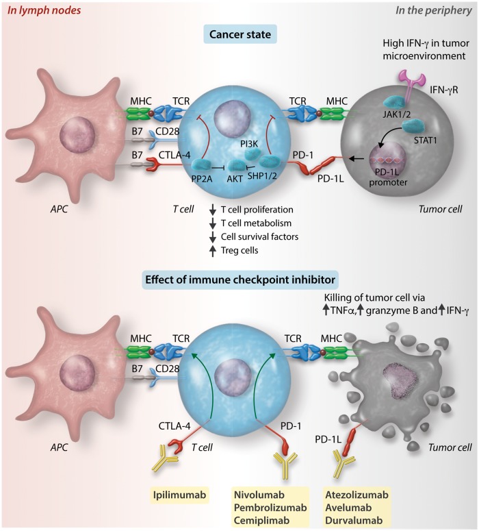
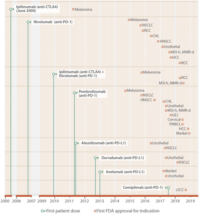
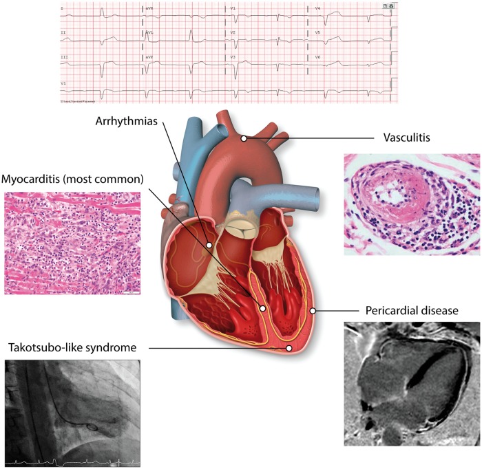
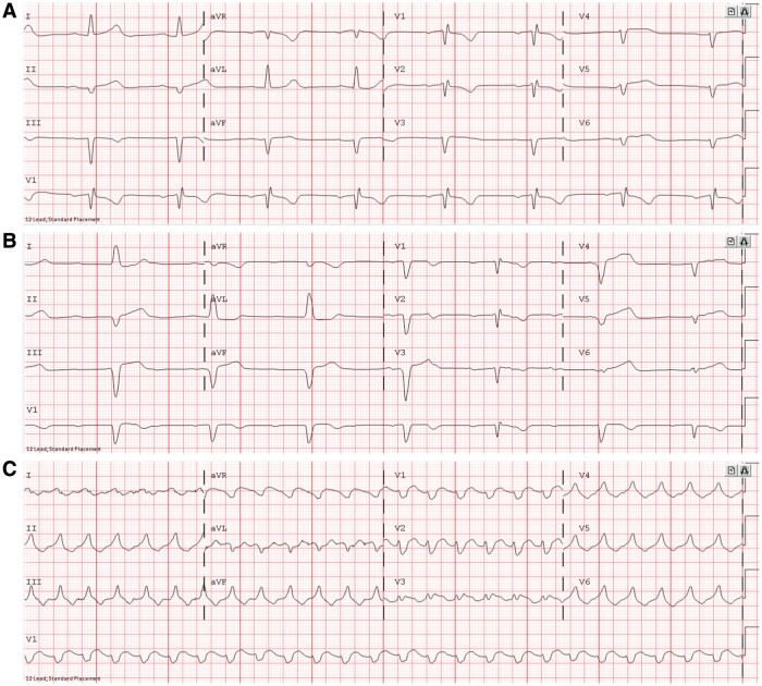
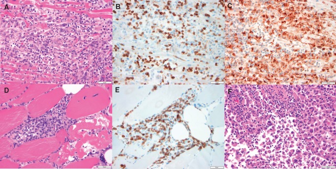
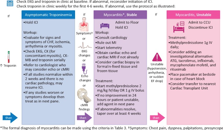
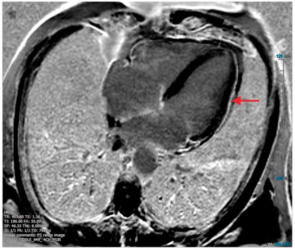
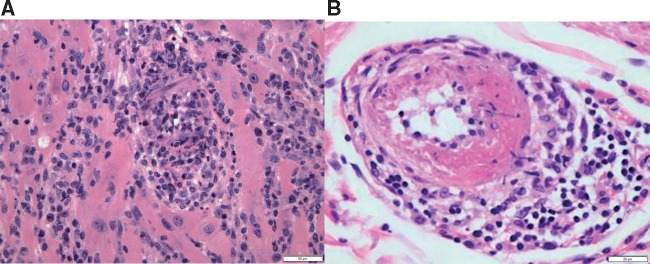
Cardiovascular toxicities associated with immune checkpoint inhibitors (ICIs) have been reported in case series but have been underappreciated due to their recent emergence, difficulties in diagnosis and non-specific clinical manifestations. ICIs are antibodies that block negative regulators of the T cell immune response, including cytotoxic T lymphocyte-associated protein-4 (CTLA-4), programmed cell death protein-1 (PD-1), and PD-1 ligand (PD-L1). While ICIs have introduced a significant mortality benefit in several cancer types, the augmented immune response has led to a range of immune-related toxicities, including cardiovascular toxicity. ICI-associated myocarditis often presents with arrhythmias, may co-exist with myositis and myasthenia gravis, can be severe, and portends a poor prognosis. In addition, pericardial disease, vasculitis, including temporal arteritis, and non-inflammatory heart failure, have been recently described as immune-related toxicities from ICI. This narrative review describes the epidemiology, diagnosis, pathophysiology, and treatment of cardiovascular toxicities of ICI therapy, highlighting recent developments in the field in the past year.

Keywords: Cardio-oncology; Cardiovascular toxicity; Immune checkpoint inhibitors; Myocarditis; Pericarditis; Vasculitis.


Figures

Figure 1
Figure 1
Cell surface receptors and ligands at immune checkpoints. Neoantigens from cancer cells are captured by APCs such as dendritic cells and presented on MHC molecules. CD8+ T cells and CD4+ T cells recognize the neoantigen-MHC I and -MHC II complexes, respectively, in the lymph node (left half of figure), become activated and migrate to the tumour bed (right half of figure). Top left: The process of T cell activation can be modulated by co-stimulatory signals from binding of B7 with CD28. Alternatively, it can be modulated by co-inhibitory signals from the binding between cytotoxic T lymphocyte-associated protein-4 (CTLA-4) and programmed cell death protein-1 (PD-1) with their respective ligands, B7 and PD-1 ligand 1 (PD-L1). Binding of CTLA-4 with B7 inhibits RACα serine/threonine-protein kinase (AKT) via activation of type II serine threonine phosphatase 2A (PP2A). Binding of PD-1 with PD-L1 or PD-L2 leads to AKT inhibition via SRC homology 2 domain-containing tyrosine phosphatase 1 and 2 (SHP1 and SHP2) inhibition of phosphoinositide 3-kinase (PI3K). Inhibition of AKT results in a range of downstream effects including reduced effector T cell proliferation, T cell metabolism, cell survival factors and increased regulatory T cell proliferation. Top right: Tumour cells can detect high levels of interferon-γ (IFN-γ) in the tumour environment via IFN-γ receptors (IFN-γR). IFN-γR signals through Janus kinase (JAK) 1 and 2, which phosphorylate and thereby activate signal transducers and activators of transcription (STAT), which dimerize and lead to promotion of transcription of PD-L1. Increased cell surface expression of PD-L1 on tumour cells enacts a ‘brake’ on the ability of T cells to mount an attack on the tumour cells. This ‘brake’ can be released by a class of anti-tumour drugs known as ICIs. Bottom: Immune checkpoints can be blocked by monoclonal antibodies against CTLA-4 (ipilimumab), PD-1 (nivolumab, pembrolizumab, and cemiplimab) and PD-L1 (atezolizumab, avelumab, and durvalumab). Blocking of these immune checkpoints restores the T cell immune response against tumour cells. Tumour necrosis factor-α, granzyme B, and interferon-γ are released, resulting in tumour cell death.
Figure 2
Figure 2
Approved ICIs and indications. Monoclonal antibodies against CTLA-4 (ipilimumab), PD-1 (nivolumab, pembrolizumab, and cemiplimab) and PD-L1 (atezolizumab, avelumab, and durvalumab) have been approved by the FDA to treat patients with a variety of advanced and metastatic cancers. The period of clinical development is illustrated from the date of the first patient dosed (green dot), and until FDA approval for specific indications (brown dot). Each month of the year is represented by a vertical line (yellow). This figure extends earlier work by Ribas and Wolchok with indications approved by the FDA in 2018. RCC, renal cell carcinoma; MSI-H, high microsatellite instability; MMR-D, mismatch repair deficient; NSCLC, non-small cell lung cancer; CHL, classical Hodgkin’s lymphoma; HNC, head and neck cancer; CRC, colorectal cancer; HCC, hepatocellular carcinoma; GEJ, gastroesophageal junction; PMBCL, primary mediastinal large B cell lymphoma; SqCC, squamous cell carcinoma.
Figure 3
Figure 3
Cardiovascular toxicity from ICIs. Cardiovascular toxicity from ICIs can come in the form of myocarditis, pericardial disease, or vasculitis. These presentations are almost never overlapping. Less commonly, it can also come in the form of Takotsubo-like syndrome and other non-inflammatory left-ventricular dysfunction. Angiography of Takotsubo-like syndrome reproduced with permission from Elsevier from Anderson and Brooks.
Figure 4
Figure 4
Electrocardiogram findings from ICI-associated myocarditis. In one patient, ECG initially showed (A) sinus bradycardia with first degree atrioventricular block, leading to (B) Sinus bradycardia with complete atrioventricular block and left bundle branch block, leading to (C) ventricular tachycardia, resulting in death. The patient was a 65-year-old woman who received one dose of nivolumab combination therapy with ipilimumab.
Figure 5
Figure 5
Histologic findings from ICI-associated myocarditis. (A–C) Photomicrographs of the myocardium, specifically, the interventricular septum. (A) Dense mononuclear cell infiltrate with extensive myocyte damage and necrosis, consistent with lymphocytic myocarditis, H&E stained section. (B) Abundance of CD3-positive T cells, CD3 immunohistochemical stain. (C) Prominent CD68-positive macrophage infiltrate, CD68 immunohistochemical stain. (D–E) are photomicrographs of skeletal muscle. (D) Dense mononuclear cell infiltrate and evidence of myocyte necrosis and damage with nuclear internalization, demonstrating myositis, H&E stained section. (E) Abundance of CD-3 positive T cells, CD3 immunohistochemical stain. (F) Photomicrograph of metastatic focus of melanoma with a lymphocytic infiltrate, H&E stained section. All histology displayed at 40× original magnification. All scale bars in this figure denote 50 µm. The patient was a 65-year-old woman who received one dose of nivolumab combination therapy with ipilimumab.
Figure 6
Figure 6
Management of ICI-associated myocarditis at Vanderbilt University Medical Center. Depicted is the screening and surveillance algorithm used at Vanderbilt for patients with increased risk of developing ICI-associated myocarditis, namely, patients on combination immunotherapy. ATG, anti-thymocyte globulin; CCU, coronary care unit; CHF, congestive heart failure; CK, creatine kinase; CK-MB, creatine kinase-muscle/brain; cMRI, cardiac magnetic resonance imaging; EKG, electrocardiogram; ICI, immune checkpoint inhibitor; IV, intravenous; MI, myocardial infarction. Icons from Servier Medical Art and the Noun Project are shared under a Creative Commons Attribution 3.0 Unported License.
Figure 7
Figure 7
Imaging findings from ICI-associated pericardial disease. cMRI of the heart demonstrating focal myocardial delayed gadolinium enhancement in the mid-lateral wall and mild late enhancement of the pericardium along the lateral wall suggestive of myopericarditis. The patient is a 70-year-old male who had received six doses enoblituzumab (an IgG antibody against B7-H3).
Figure 8
Figure 8
Histologic findings from ICI-associated vasculitis. (A) Photomicrograph of myocardium with lymphocytic myocarditis and vasculitis, with obliteration of the lumen of this small artery. H&E stained section, 200× original magnification, with scale bar denoting 50 µm. (B) Photomicrograph of small artery with fibrinoid necrosis and perivascular chronic inflammation. H&E stained section, 400× original magnification, with scale bar denoting 20 µm.

References

    1. Farkona S, Diamandis EP, Blasutig IM.. Cancer immunotherapy: the beginning of the end of cancer? BMC Med 2016;14:73. - PMC - PubMed
    1. Ribas A, Wolchok JD.. Cancer immunotherapy using checkpoint blockade. Science 2018;359:1350–1355. - PMC - PubMed
    1. Taube JM, Anders RA, Young GD, Xu H, Sharma R, McMiller TL, Chen S, Klein AP, Pardoll DM, Topalian SL, Chen L.. Colocalization of inflammatory response with B7-H1 expression in human melanocytic lesions supports an adaptive resistance mechanism of immune escape. Sci Transl Med 2012;4: 127ra37. - PMC - PubMed
    1. Johnson DB, Chandra S, Sosman JA.. Immune checkpoint inhibitor toxicity in 2018. JAMA 2018;320:1702–1703. - PubMed
    1. Friedman CF, Proverbs-Singh TA, Postow MA.. Treatment of the immune-related adverse effects of immune checkpoint inhibitors: a review. JAMA Oncol 2016;2:1346–1353. - PubMed

Publication types

Substances