. 2019 May 7;47(8):4026-4038.
doi: 10.1093/nar/gkz056.

PrimPol is required for the maintenance of efficient nuclear and mitochondrial DNA replication in human cells

Affiliations

PrimPol is required for the maintenance of efficient nuclear and mitochondrial DNA replication in human cells

Laura J Bailey et al. Nucleic Acids Res. .

Abstract

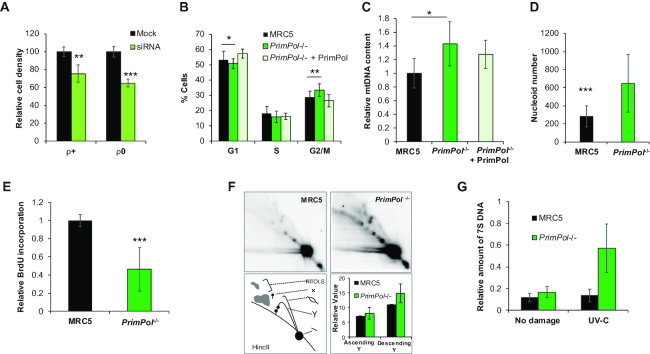
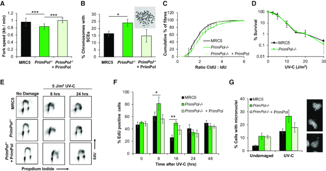
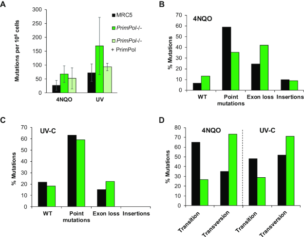
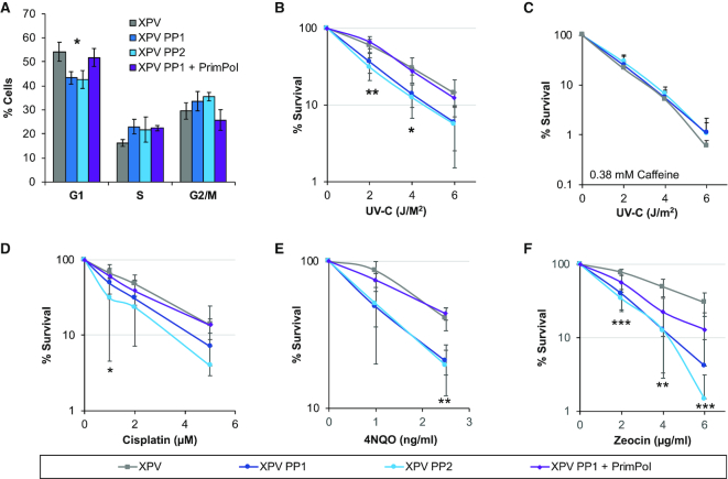
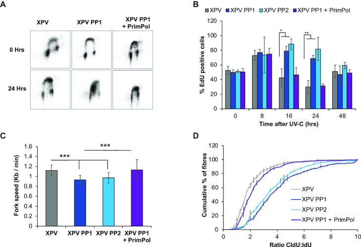
Eukaryotic Primase-Polymerase (PrimPol) is an enzyme that maintains efficient DNA duplication by repriming replication restart downstream of replicase stalling lesions and structures. To elucidate the cellular requirements for PrimPol in human cells, we generated PrimPol-deleted cell lines and show that it plays key roles in maintaining active replication in both the nucleus and mitochondrion, even in the absence of exogenous damage. Human cells lacking PrimPol exhibit delayed recovery after UV-C damage and increased mutation frequency, micronuclei and sister chromatin exchanges but are not sensitive to genotoxins. PrimPol is also required during mitochondrial replication, with PrimPol-deficient cells having increased mtDNA copy number but displaying a significant decrease in replication. Deletion of PrimPol in XPV cells, lacking functional polymerase Eta, causes an increase in DNA damage sensitivity and pronounced fork stalling after UV-C treatment. We show that, unlike canonical TLS polymerases, PrimPol is important for allowing active replication to proceed, even in the absence of exogenous damage, thus preventing the accumulation of excessive fork stalling and genetic mutations. Together, these findings highlight the importance of PrimPol for maintaining efficient DNA replication in unperturbed cells and its complementary roles, with Pol Eta, in damage tolerance in human cells.


Figures

Figure 1.
Figure 1.
PrimPol knockout cells proliferate normally, have increased mtDNA content but exhibit decreased replication rates. (A) Human Osteosarcoma cells (143B) treated with PrimPol siRNA or scrambled control were monitored using an IncuCyte™ microscope for 80 h with cells imaged every 3 h and cell density calculated (see Supplementary Figure S1). This shows the mean cell density at 60 h after RNAi from n = 3 experiments with standard deviation shown with error bars. (B) Cell cycle populations were measured in asynchronously growing MRC5 and PrimPol−/− cells or PrimPol−/− cells complemented with GFP-tagged PrimPol after PI staining and flow cytometry. (C) mtDNA copy number was analysed by Taqman probe qPCR in human MRC5 cells with and without PrimPol. (D) The mitochondrial network and mtDNA nucleoids were analysed by co-staining cells with Mitotracker (red), anti-DNA (green) and DAPI (blue) and ImageJ was used to analyse the number of mtDNA nucleoids per cell. (E) mtDNA replication was measured using BrdU incorporation, nuclear DNA replication was blocked using aphidicolin to allow mtDNA-specific analysis and cells were allowed to accumulate BrdU for 24 h. DNA was extracted, dot blotted and BrdU content quantified by anti-BrdU staining, this was then compared to total DNA content by the use of a mtDNA-specific radio-labelled probe, n = 3 independent repeats performed in triplicate. (F) mtDNA replication intermediates were viewed directly by 2D-AGE, total DNA was digested by HinCII and separated in two dimensions. DNA was Southern blotted and probed with an OH specific radio-labelled probe. Images were quantified using AIDA. (G) 7S DNA copy number was measured in undamaged and UV-C treated cells using southern blotting of whole cell DNA after PvuII digestion and probed with a 7S DNA and nuclear DNA specific radio-labelled probe, and quantified using AIDA software. Charts represent the mean of n≥3 independent experiments with error bars showing standard deviation, significance was measured using a Student's t-test, *P≤ 0.05, **P≤ 0.01, ***P≤ 0.001.
Figure 2.
Figure 2.
PrimPol−/− cells have decreased replication speeds, increased damage recovery times and micronuclei. (A) Replication fork speeds were measured in WT and PrimPol−/− cells or PrimPol−/− complemented with GFP-PrimPol by labelling forks with CldU, followed by IdU and measuring individual fibres after spreading. 300 fibres were measured over 3 independent experiments. P values were 2.06 × 10⁻¹⁰ for MRC5:PrimPol−/−, 0.236 for MRC5:PrimPol−/− + PrimPol and 1.24 × 10⁻¹⁴ for PrimPol−/−:PrimPol−/−+ PrimPol. (B) Percentage of chromosomes carrying one or more sister chromatid exchanges (SCEs) were counted. Data represents three independent experiments, where 200+ chromosomes were analysed per slide. Error bars show standard deviation and a representative image of WT cells is shown above. (C) This figure shows the accumulated percentage of ratios of the two labels, where a pulse of 20 J/m2 UV-C was given in between the two labels. Data is shown as the average across three independent experiments (∼300 fibres) with error bars representing standard deviation. Student's t-test on this data showed P values of 0.0000106 for MRC5: PrimPol−/− (*** significance), 0.09 for MRC5: PrimPol−/− + PrimPol (not significantly different) and 0.0024 for PrimPol−/− + PrimPol (** significance). (D) Colony survival assays were used to compare the sensitivity of cells to UV-C damage, chart represents n ≥ 3 independent repeats with error bars showing standard deviation. (E) WT and PrimPol−/− cells or PrimPol−/− complemented with GFP-PrimPol were labelled with EdU at increasing time points after 5 J/m2 UV-C damage. After being fixed and dual labelled with propidium iodide, cells were analysed by flow cytometry. (F) This shows the percentage of EdU positive cells in early S-phase at increasing times after damage n ≥ 3, error bars show standard deviation. (G) Cells were stained with DAPI 72 h after 0 or 5 J/m2 UV-C damage and number of cells with one or more micronucleus were counted as a percentage of the total cell population, charts show the mean of three or more independent experiments with standard deviation shown by error bars. Representative images are shown on the right. Significance was measured using a Student's t-test, * P≤ 0.05, ** P≤ 0.01, *** P≤ 0.001.
Figure 3.
Figure 3.
PrimPol−/− cells have increased mutation frequency after damage. Mutation frequencies were calculated after 5 days following 40 ng/ml 4NQO treatment or 10 J/m2 UV-C damage, followed by 6–7 days recovery. Cells were plated in the presence of 5 μg/ml 6-thioguanine to identify clones with a mutation in the HPRT gene and the number of colonies were calculated compared to a drug free control plate. (A) Mutation frequency was then calculated as occurrence of a mutation per 106 cells, bars show the average of ≥3 independent experiments and error bars represent standard deviation. RNA was extracted from 6-Thioguanine-resistant clones and the HPRT gene cDNA was amplified and sequenced to identify mutations generated after 4NQO treatment (B) or UV-C damage (C), shown as a percentage of the whole population. Details of the identified clones shown in Supplementary Figure S4F. (D) The percentage of point mutations due to transitions or transversions in MRC5 and PrimPol−/− cells after treatment with UV-C or 4NQO. P = 0.0086 using two-way ANOVA.
Figure 4.
Figure 4.
Loss of both PrimPol and Pol η leads to greater damage sensitivity. (A) Cell cycle population changes were studied in WT (XPV) and PrimPol−/− XP30RO cells (XPV PP1 & XPV PP2) and those complemented with GFP PrimPol (XPV PP1 + PrimPol) by flow cytometry analysis of propidium iodide labelled cells. (B–F) Sensitivity of cells to a range of DNA damaging agents was measured using colony survival assays. Cells were treated with increasing doses of UV-C in the absence or presence of 0.38 mM caffeine (B and C) or increasing doses of cisplatin for 5 h, after which the cells were washed free of the drug (D) or cells were maintained in media containing increasing doses of 4NQO or zeocin (E and F) for 10 days to form colonies before being stained with methylene blue and counted. Charts represent the mean of n ≥ 3 independent experiments with error bars showing standard deviation. Significance was measured using a Student's t-test, *P≤ 0.05, **P≤ 0.01, ***P≤ 0.001.
Figure 5.
Figure 5.
XPV PP cells have an increase in replication defects. Cells were treated with 2 J/m2 UV-C and allowed to recover for increasing times before being labelled with EdU. Cell cycle populations were then measured by the addition of propidium iodide, representative images are shown in (A) with percentage of EdU cells in early S-phase quantified in (B). Replication fork speeds were measured in undamaged cells by labelling with CldU followed by IdU, after spreading and staining with specific antibodies, fibres were measured using OMERO, 300 fibres were measured over 3 independent experiments (C). P values were 1.33 × 10⁻²⁰ for XPV: XPV PP1, 1.91 × 10⁻¹⁴ for XPV: XPV PP2, 0.849 for XPV: XPV PP1 + PrimPol and 1.1 × 10⁻¹³ for XPV PP1: XPV PP1 + PrimPol. Fork stalling was measured by the addition of a 20 J/m2 pulse of UV-C, between the addition of the two labels, and the ratio of the two labels was compared and is shown as the accumulated percentage of the average of each experiment (D). P values for the raw data were 5.89 × 10⁻²⁶ for XPV: XPV PP1, 2.51 × 10⁻²⁰ for XPV: XPV PP2, 0.273 for XPV: XPV PP1 + PrimPol and 1.74 × 10⁻²² for XPV PP1: XP PP1 + PrimPol. Charts represent the mean of n ≥ 3 independent experiments with error bars showing standard deviation. Significance was measured using a Student's t-test, *P≤ 0.05, **P≤ 0.01, ***P≤ 0.001.

References

    1. Iyama T., Wilson D.M. 3rd.. DNA repair mechanisms in dividing and non-dividing cells. DNA Repair (Amst.). 2013; 12:620–636. - PMC - PubMed
    1. Vaisman A., Woodgate R.. Translesion DNA polymerases in eukaryotes: what makes them tick. Crit. Rev. Biochem. Mol. Biol. 2017; 52:274–303. - PMC - PubMed
    1. Trakselis M.A., Cranford M.T., Chu A.M.. Coordination and substitution of DNA polymerases in response to genomic obstacles. Chem. Res. Toxicol. 2017; 30:1956–1971. - PubMed
    1. Sale J.E., Lehmann A.R., Woodgate R.. Y-family DNA polymerases and their role in tolerance of cellular DNA damage. Nat. Rev. Mol. Cell Biol. 2012; 13:141–152. - PMC - PubMed
    1. Livneh Z., Ziv O., Shachar S.. Multiple two-polymerase mechanisms in mammalian translesion DNA synthesis. Cell Cycle. 2010; 9:729–735. - PubMed

Publication types

MeSH terms