. 2019 May;33(5):6129-6139.
doi: 10.1096/fj.201802388R. Epub 2019 Feb 4.

TBC1D3 regulates the payload and biological activity of extracellular vesicles that mediate tissue repair

Affiliations

TBC1D3 regulates the payload and biological activity of extracellular vesicles that mediate tissue repair

Shu Qin et al. FASEB J. 2019 May.

Abstract

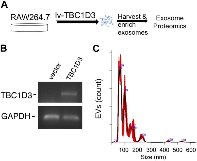
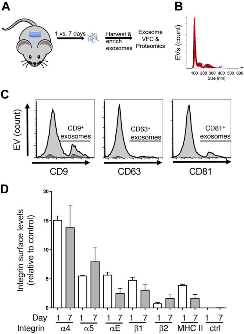
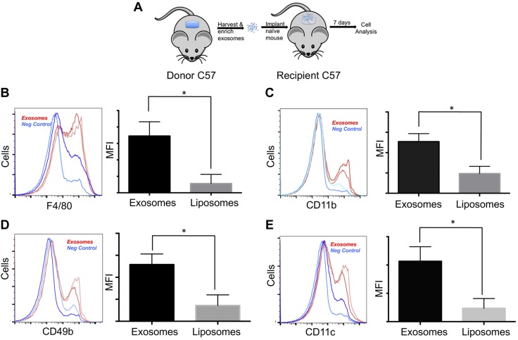
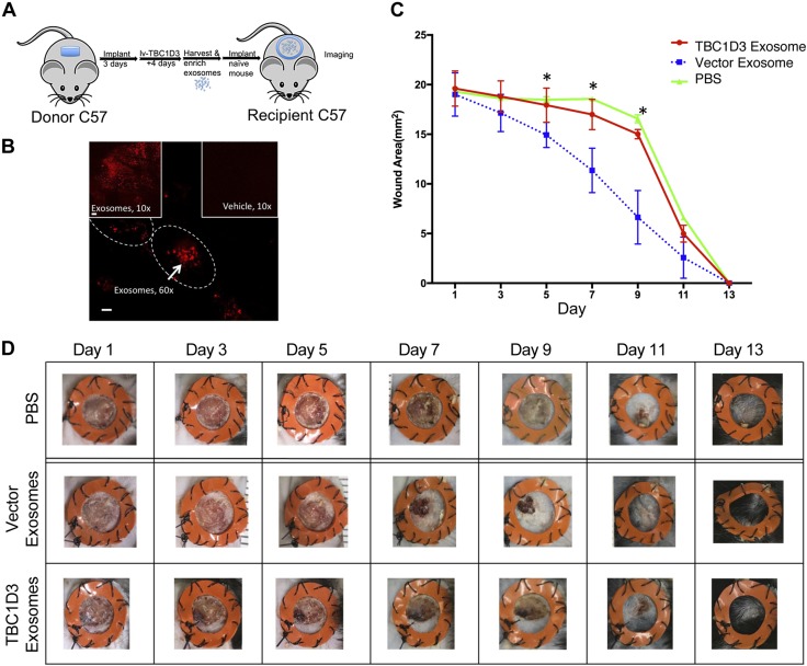
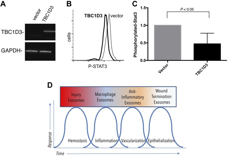
Healthy repair of cutaneous injury is a coordinated response of inflammatory cells, secreted factors, and biologically active extracellular vesicles (EVs). Although constitutive release of EVs into biologic fluids is a hallmark of cultured cells and tumors, their payload and biologic activity appears to be tightly regulated. We show that Tre-2/Bub2/Cdc16 (TBC1) domain family member 3 (TBC1D3) drives the release of an EV population that causes a decrease in phosphorylation of the transcription factor signal transducer and activator of transcription 3 in naive recipient cells. To explore the biologic activity of EVs in vivo, we used a mouse model of sterile subcutaneous inflammation to determine the payload and biologic activity of EVs released into the microenvironment by committed myeloid lineages and stroma. Expression of TBC1D3 in macrophages altered the payload of their released EVs, including RNA-binding proteins, molecular motors, and proteins regulating secretory pathways. A wound-healing model demonstrated that closure was delayed by EVs released under the control of TBC1D3. We show that modulating the secretory repertoire of a cell regulates EV payload and biologic activity that affects outcomes in tissue repair and establishes a strategy for modifying EVs mediating specific biologic responses.-Qin, S., Dorschner, R. A., Masini, I., Lavoie-Gagne, O., Stahl, P. D., Costantini, T. W., Baird, A., Eliceiri, B. P. TBC1D3 regulates the payload and biological activity of extracellular vesicles that mediate tissue repair.

Keywords: RabGAP; exosome; foreign body response; macrophage activation; wound healing.


Conflict of interest statement

The authors thank Ann-Marie Hageny [University of California–San Diego (UCSD)] for expert technical support, and Dr. Antonio DeMaio (UCSD) for use of the Nanoparticle Tracking Analysis Instrument; Dr. Majid Ghassemian (UCSD Biomolecular and Proteomics Mass Spectrometry Facility) provided expert support and analysis for the proteomics studies; Dr. John Nolan (Scintillon Institute, San Diego, CA, USA) provided expert advice on the design of vesicle flow cytometry assays; Dr. Chen Kong (Washington University) helped with consultation on the molecular biology of TBC1D3; and Dr. Lisa A. Hannan provided expert editorial review of the study. Research was supported by operating grants from the U.S. National Institutes of Health, National Institute of General Medical Sciences (1R01GM121530), the UCSD Academic Senate, the UCSD Department of Surgery Reinvestment Fund, the Shock Society, and the Surgical Infection Society. The authors declare no conflicts of interest.

Figures

Figure 1
Figure 1
Expression of TBC1D3 in macrophages regulates the payload of released EVs. A) Mouse RAW264.7 macrophages were transduced with lentivirus-expressing TBC1D3 or vector control. B) The cells validated for expression of TBC1D3 by RT-PCR. C) EVs isolated and subjected to NTA with a representative plot shown from vector-transduced cells. No significant differences were observed in the NTA between vector and TBC1D3-transduced RAW264.7 cell EVs.
Figure 2
Figure 2
Kinetics of EV release defined by changes in protein profile and payload in the foreign-body response in vivo. A) Subcutaneous implants of PVA sponge were used as a model for sterile inflammation where EVs are isolated from the conditioned supernatant of infiltrating leukocytes. B) Representative NTA analysis of d 7 EVs showing a mode value of 144.9 ± 51, with no significant differences with d 1 EVs. C) VFC analysis was performed on vesicles released in the PVA sponge implant after 7 d using antibodies directed to the tetraspanins CD9, CD63, and CD81 and are indicated under the line segment (light gray). Treatment of samples with the detergent Triton X-100 was used to establish background values (dark gray shading). D) VFC analysis of integrin subunits and major histocompatibility complex (MHC) II detected on EVs collected at d 7 postimplantation to assess the profile of integrins present on EVs. No significant differences in integrins were observed between d 1 and d 7 EVs. Ctrl, control.
Figure 3
Figure 3
Adoptive transfer of EVs from 7 d donor site promotes macrophage recruitment in vivo. A) Adoptive transfer of EVs from donor site PVA sponge implants that are then purified and adoptively transferred to naive PVA sponge implants to monitor leukocyte recruitment. BE) Flow cytometry analysis of leukocytes infiltrating recipient PVA sponges that had received EVs isolated from d 7 donor PVA implants, incubated for an additional 7 d post-transfer. EVs (red lines) are compared to liposome controls (blue lines) from duplicate samples. For each group, an analysis of F4/80 levels by histogram and quantitation of mean fluorescence intensity (MFI) is provided (B), along with an analysis of CD11b+ cells by histogram and MFI (C), CD49b+ cells by histogram and MFI (D), and CD11c+ cells by histogram and MFI (E). For each marker, n = 2. *P < 0.05.
Figure 4
Figure 4
Expression of TBC1D3 regulates the biologic activity of a class of EVs that uncouples the proreparative effects of EVs on wound closure. A) Overview of the lentiviral transduction of leukocytes infiltrating the PVA donor site from which EVs are isolated for adoptive transfer of EVs into recipient 4-mm full-thickness cutaneous punches. B) Donor EVs labeled with fluorescent PKH26 were transferred to fresh wound sites, incubated for 18 h, imaged by confocal microscopy, and compared with vehicle controls. Cell membrane is indicated with a tracing to show intracellular clustering of EV uptake. Scale bar, 60 µm. C) Quantification of the kinetics of wound closure was determined in splinted wound sites following the addition of EVs harvested from the PVA donor site cells expressing TBC1D3 (red) vs. EVs isolated from vector control cells (blue). PBS was used as a control for the normal kinetics of wound closure in the absence of EVs (green). *P < 0.05 between TBC1D3 and vector groups; n = 6 for each group. D) Representative images of the time course of wound closure following addition of TBC1D3-regulated EVs, vector control EVs, and buffer control.
Figure 5
Figure 5
TBC1D3 expression in human hematopoietic and myeloid cells. A) RT-PCR validation of TBC1D3 gene delivery in THP-1 cells. B) Conditioned medium of donor TBC1D3-transduced cells was enriched by ultracentrifugation and added to naive recipient THP-1 cells for 18 h, fixed, permeabilized, and stained for intracellular levels of phosphorylated STAT3 compared with vector control. C) Quantification of TBC1D3-mediated changes on the biologic activity of EVs released under the regulation of TBC1D3 on the phosphorylation of STAT3 (P < 0.05, n = 3). D) Model for the role of EVs in defining phases of wound healing based on the hypothesis that EV classes are dependent on the kinetics of tissue repair.

References

    1. Kojima M., Gimenes-Junior J. A., Langness S., Morishita K., Lavoie-Gagne O., Eliceiri B., Costantini T. W., Coimbra R. (2017) Exosomes, not protein or lipids, in mesenteric lymph activate inflammation: unlocking the mystery of post-shock multiple organ failure. J. Trauma Acute Care Surg. 82, 42–50 - PubMed
    1. Lee H., Zhang D., Zhu Z., Dela Cruz C. S., Jin Y. (2016) Epithelial cell-derived microvesicles activate macrophages and promote inflammation via microvesicle-containing microRNAs. Sci. Rep. 6, 35250 - PMC - PubMed
    1. Li D., Liu J., Guo B., Liang C., Dang L., Lu C., He X., Cheung H. Y., Xu L., Lu C., He B., Liu B., Shaikh A. B., Li F., Wang L., Yang Z., Au D. W., Peng S., Zhang Z., Zhang B. T., Pan X., Qian A., Shang P., Xiao L., Jiang B., Wong C. K., Xu J., Bian Z., Liang Z., Guo D. A., Zhu H., Tan W., Lu A., Zhang G. (2016) Osteoclast-derived exosomal miR-214-3p inhibits osteoblastic bone formation. Nat. Commun. 7, 10872 - PMC - PubMed
    1. Shenoda B. B., Ajit S. K. (2016) Modulation of immune responses by exosomes derived from antigen-presenting cells. Clin. Med. Insights Pathol. 9 (Suppl 1), 1–8 - PMC - PubMed
    1. Garzetti L., Menon R., Finardi A., Bergami A., Sica A., Martino G., Comi G., Verderio C., Farina C., Furlan R. (2014) Activated macrophages release microvesicles containing polarized M1 or M2 mRNAs. J. Leukoc. Biol. 95, 817–825 - PubMed

Publication types

Substances

LinkOut - more resources