. 2019 Feb 4;14(2):e0211647.
doi: 10.1371/journal.pone.0211647. eCollection 2019.

Molecular insights in the pathogenesis of classical Ehlers-Danlos syndrome from transcriptome-wide expression profiling of patients' skin fibroblasts

Affiliations

Molecular insights in the pathogenesis of classical Ehlers-Danlos syndrome from transcriptome-wide expression profiling of patients' skin fibroblasts

Nicola Chiarelli et al. PLoS One. .

Abstract

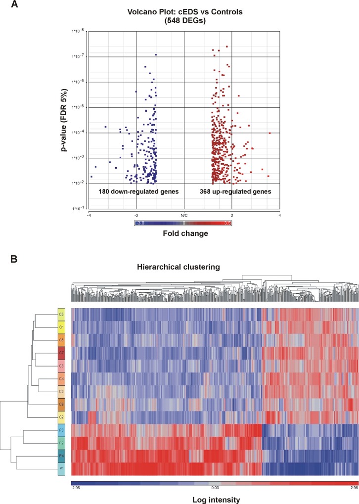
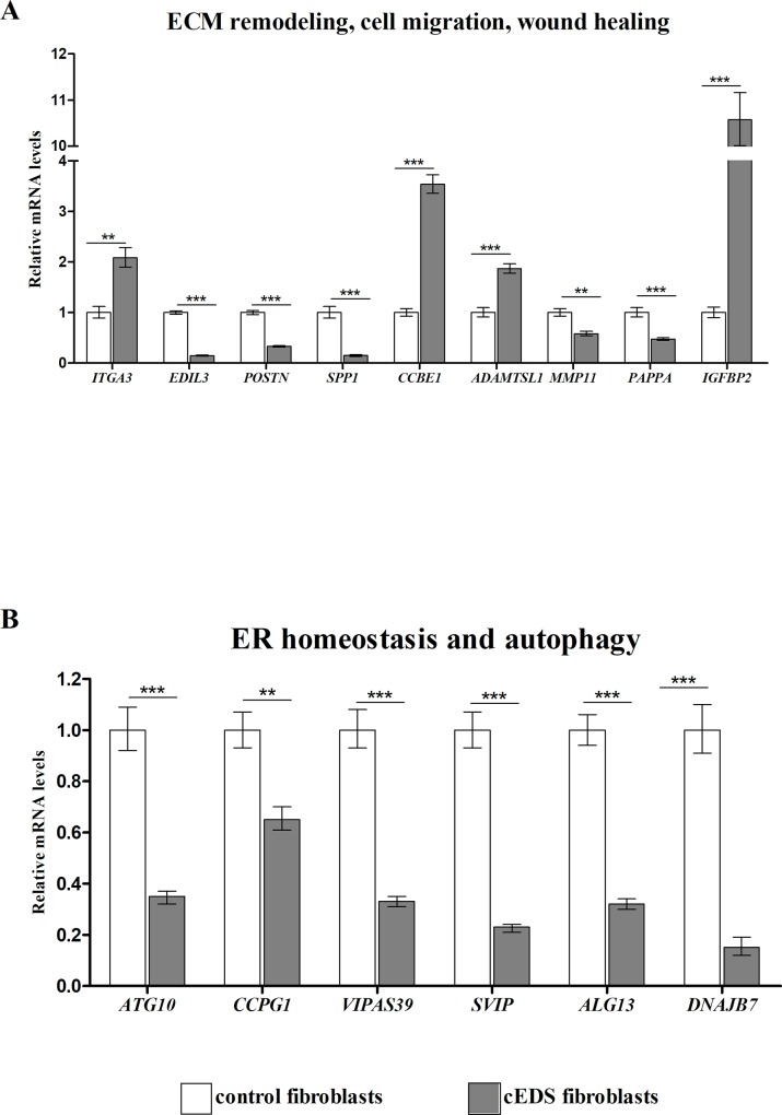
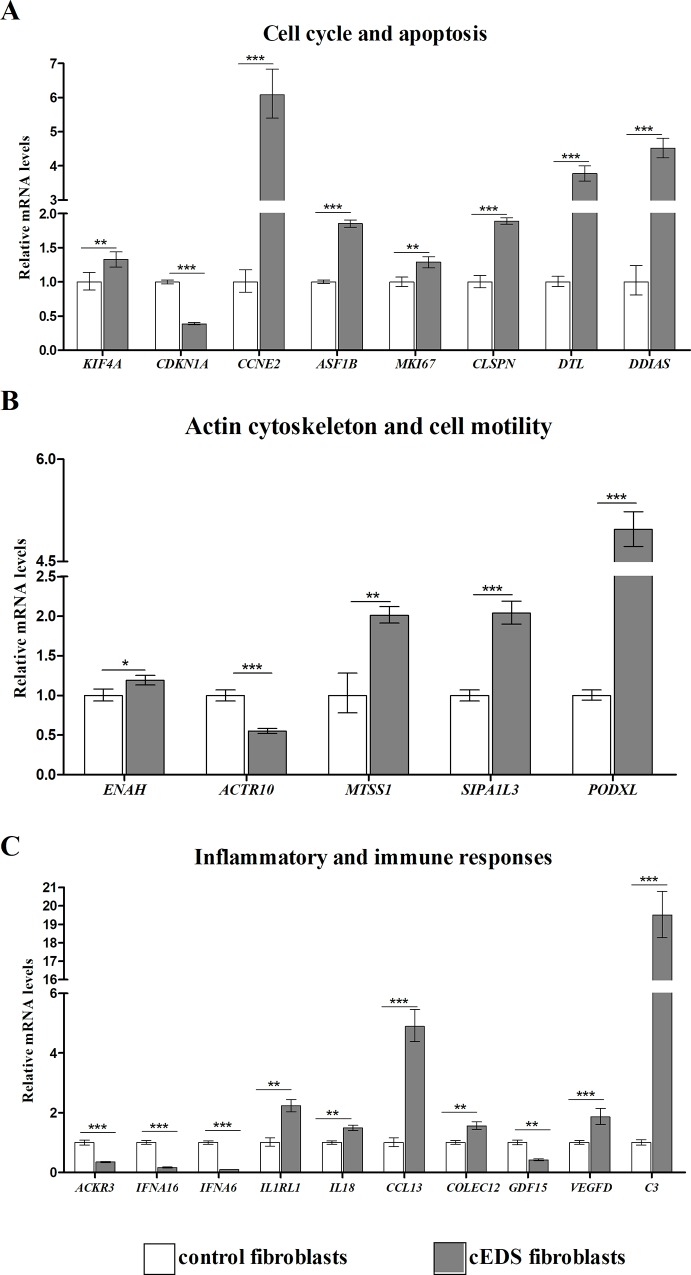
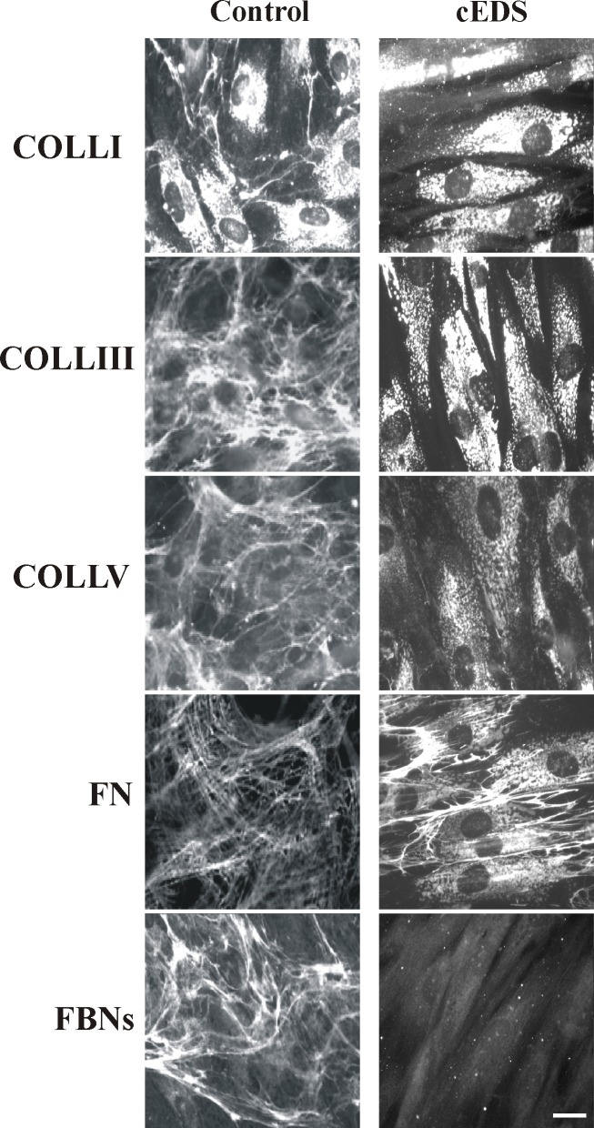
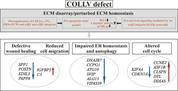
Classical Ehlers-Danlos syndrome (cEDS) is a dominant inherited connective tissue disorder mainly caused by mutations in the COL5A1 and COL5A2 genes encoding type V collagen (COLLV), which is a fibrillar COLL widely distributed in a variety of connective tissues. cEDS patients suffer from skin hyperextensibility, abnormal wound healing/atrophic scars, and joint hypermobility. Most of the causative variants result in a non-functional COL5A1 allele and COLLV haploinsufficiency, whilst COL5A2 mutations affect its structural integrity. To shed light into disease mechanisms involved in cEDS, we performed gene expression profiling in skin fibroblasts from four patients harboring haploinsufficient and structural mutations in both disease genes. Transcriptome profiling revealed significant changes in the expression levels of different extracellular matrix (ECM)-related genes, such as SPP1, POSTN, EDIL3, IGFBP2, and C3, which encode both matricellular and soluble proteins that are mainly involved in cell proliferation and migration, and cutaneous wound healing. These gene expression changes are consistent with our previous protein findings on in vitro fibroblasts from other cEDS patients, which exhibited reduced migration and poor wound repair owing to COLLV disorganization, altered deposition of fibronectin into ECM, and an abnormal integrin pattern. Microarray analysis also indicated the decreased expression of DNAJB7, VIPAS39, CCPG1, ATG10, SVIP, which encode molecular chaperones facilitating protein folding, enzymes regulating post-Golgi COLLs processing, and proteins acting as cargo receptors required for endoplasmic reticulum (ER) proteostasis and implicated in the autophagy process. Patients' cells also showed altered mRNA levels of many cell cycle regulating genes including CCNE2, KIF4A, MKI67, DTL, and DDIAS. Protein studies showed that aberrant COLLV expression causes the disassembly of itself and many structural ECM constituents including COLLI, COLLIII, fibronectin, and fibrillins. Our findings provide the first molecular evidence of significant gene expression changes in cEDS skin fibroblasts highlighting that defective ECM remodeling, ER homeostasis and autophagy might play a role in the pathogenesis of this connective tissue disorder.


Conflict of interest statement

The authors have declared that no competing interests exist.

Figures

Fig 1
Fig 1. Volcano plot and hierarchical clustering analyses for DEGs in skin fibroblasts from cEDS patients and control subjects.
(A) The volcano plot depicts all statistically significant 548 DEGs (180 down-regulated, 368 upregulated) identified in cEDS cells. The fold-change of DEGs on the x-axis vs the statistical significance (FDR-adjusted p-value <0.05) on the y-axis is shown; the up-regulated genes are reported in red, and the down-regulated genes are in blue. (B) Hierarchical clustering analysis of DEGs identified in cEDS fibroblasts. Although fibroblasts from only 9 control subjects (C) and 4 cEDS patients (P) were analyzed, this analysis showed the presence of two distinct clusters of transcripts that clearly distinguish the patients from the healthy individuals. The red color represents high gene expression, and blue represents low gene expression.
Fig 2
Fig 2. qPCR validation of genes related to ECM remodeling, wound healing, and ER homeostasis.
(A) The relative mRNA expression levels of selected genes related to ECM architecture and (B) involved in ER homeostasis and autophagy, were determined with the 2-(ΔΔCt) method normalized with the geometric mean of different reference genes. Bars represent the mean ratio of target gene expression in four patients’ fibroblasts compared to six unrelated healthy individuals. qPCR was performed in triplicate, and the results were expressed as mean ± SEM. **p<0.01, and ***p<0.001.
Fig 3
Fig 3. qPCR validation of genes involved in cell cycle regulation, actin cytoskeleton organization, and related to inflammatory and immune responses.
(A) The relative mRNA expression levels of different cell cycle regulating genes, (B) transcripts associated with cytoskeleton organization, and (C) implicated in inflammation and immune response, were determined with the 2-(ΔΔCt) method normalized with the geometric mean of different reference genes. Bars represent the mean ratio of target gene expression in four patients’ fibroblasts compared to six unrelated healthy individuals. qPCR was performed in triplicate, and the results were expressed as mean ± SEM. *p<0.05, **p<0.01, and ***p<0.001.
Fig 4
Fig 4. cEDS patients’ skin fibroblasts show the altered deposition of different structural components into the ECM.
Control and patients’ cells were analyzed with specific Abs directed against COLLI, COLLIII, COLLV, FN, and FBNs. FN and FBNs were investigated by labeling the cells with Abs recognizing all their isoforms. Images are representative of 4 control and 4 cEDS cell strains. Scale bar: 10 μm.
Fig 5
Fig 5. Schematic summary illustrating the main biological processes deregulated in skin fibroblasts with COL5A1 and COL5A2 pathogenetic variants.
COLLV defect is associated with pathological ECM remodeling and anoikis, which is rescued by an αvβ3 integrin-EGFR signaling transduction pathway [20], together with defective ER homeostasis, autophagy and cell cycle progression. Down-regulated DEGs are indicated by blue arrows and those up-regulated by red arrows.

References

    1. Malfait F, Francomano C, Byers P, Belmont J, Berglund B, Black J, et al. The 2017 international classification of the Ehlers-Danlos syndromes. Am J Med Genet C Semin Med Genet. 2017; 175: 8–26. 10.1002/ajmg.c.31552 - DOI - PubMed
    1. Bowen JM, Sobey GJ, Burrows NP, Colombi M, Lavallée ME, Malfait F, et al. Ehlers-Danlos syndrome, classical type. Am J Med Genet C Semin Med Genet. 2017; 175: 27–39. 10.1002/ajmg.c.31548 - DOI - PubMed
    1. Colombi M, Dordoni C, Venturini M, Ciaccio C, Morlino S, Chiarelli N, et al. Spectrum of mucocutaneous, ocular and facial features and delineation of novel presentations in 62 classical Ehlers-Danlos syndrome patients. Clin Genet. 2017; 92: 624–631. 10.1111/cge.13052 - DOI - PubMed
    1. Symoens S, Syx D, Malfait F, Callewaert B, Vanakker O, Coucke P, et al. Comprehensive molecular analysis demonstrates type V collagen mutations in over 90% patients with classical EDS and allows to refine diagnostic criteria. Hum Mutat. 2012; 33: 1485–1493. 10.1002/humu.22137 - DOI - PubMed
    1. Ritelli M, Dordoni C, Venturini M, Chiarelli N, Quinzani S, Traversa M, et al. Clinical and molecular characterization of 40 patients with classic Ehlers–Danlos syndrome: Identification of 18 COL5A1 and 2 COL5A2 novel mutations. Orphanet J Rare Dis. 2013; 8:58 10.1186/1750-1172-8-58 - DOI - PMC - PubMed

MeSH terms