. 2019 Feb 1;17(2):92.
doi: 10.3390/md17020092.

New Source of 3D Chitin Scaffolds: The Red Sea Demosponge Pseudoceratina arabica (Pseudoceratinidae, Verongiida)

Affiliations

New Source of 3D Chitin Scaffolds: The Red Sea Demosponge Pseudoceratina arabica (Pseudoceratinidae, Verongiida)

Lamiaa A Shaala et al. Mar Drugs. .

Abstract

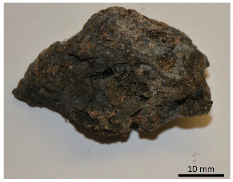
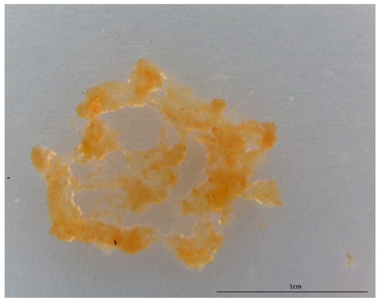
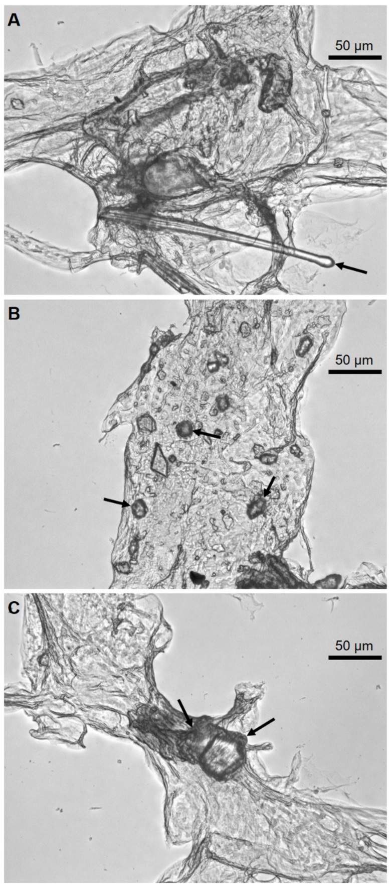
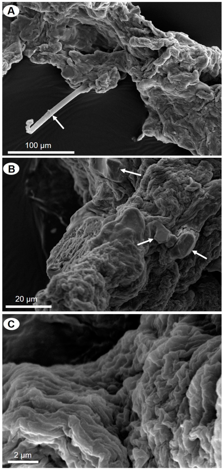
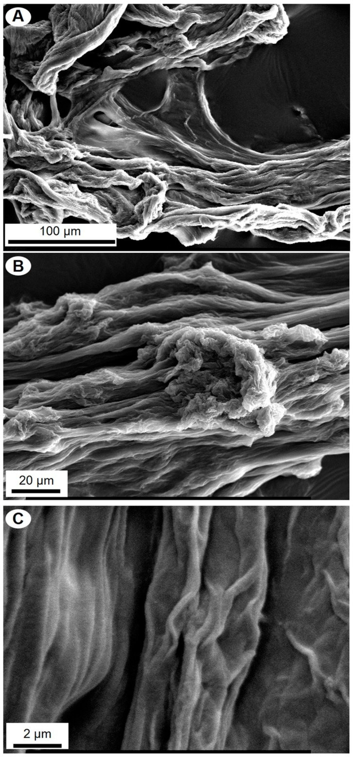
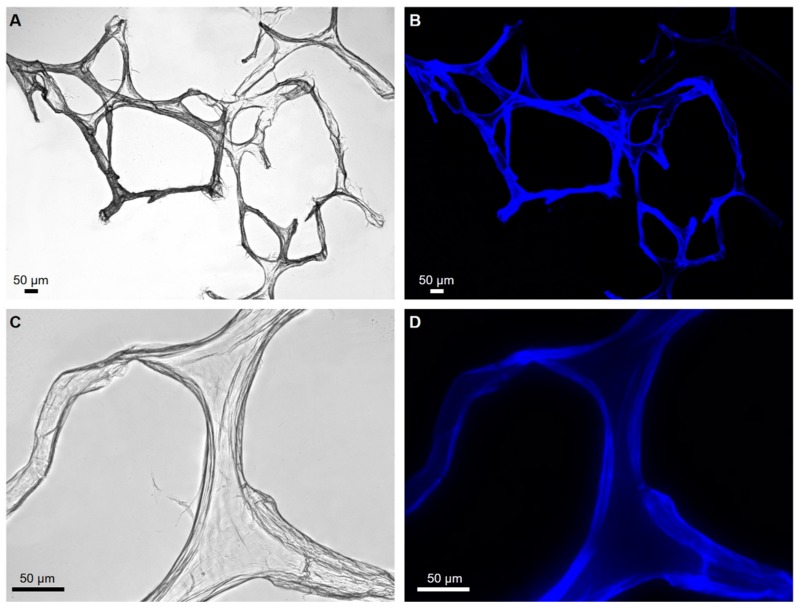
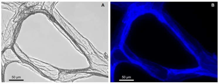
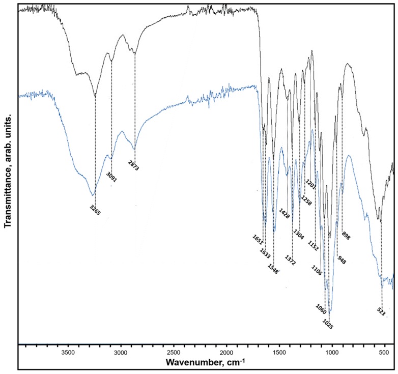
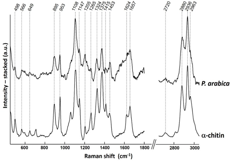
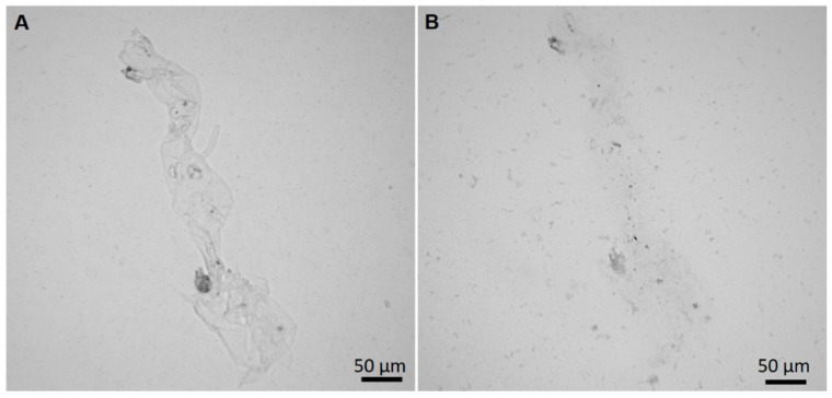
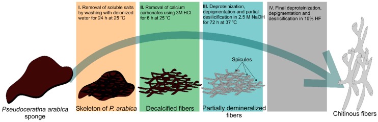
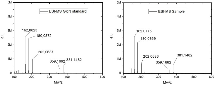
The bioactive bromotyrosine-derived alkaloids and unique morphologically-defined fibrous skeleton of chitin origin have been found recently in marine demosponges of the order Verongiida. The sophisticated three-dimensional (3D) structure of skeletal chitinous scaffolds supported their use in biomedicine, tissue engineering as well as in diverse modern technologies. The goal of this study was the screening of new species of the order Verongiida to find another renewable source of naturally prefabricated 3D chitinous scaffolds. Special attention was paid to demosponge species, which could be farmed on large scale using marine aquaculture methods. In this study, the demosponge Pseudoceratina arabica collected in the coastal waters of the Egyptian Red Sea was examined as a potential source of chitin for the first time. Various bioanalytical tools including scanning electron microscopy (SEM), fluorescence microscopy, FTIR analysis, Calcofluor white staining, electrospray ionization mass spectrometry (ESI-MS), as well as a chitinase digestion assay were successfully used to confirm the discovery of α-chitin within the skeleton of P. arabica. The current finding should make an important contribution to the field of application of this verongiid sponge as a novel renewable source of biologically-active metabolites and chitin, which are important for development of the blue biotechnology especially in marine oriented biomedicine.

Keywords: Pseudoceratina arabica; biological materials; chitin; demosponges; scaffolds.


Conflict of interest statement

The authors declare no conflict of interest.

Figures

Figure 1
Figure 1
The fragment of the dried specimens of P. arabica demosponge used in this study.
Figure 2
Figure 2
Completely demineralized and pigment-free scaffolds isolated from the sponge P. arabica.
Figure 3
Figure 3
Alkali-treated fibers of P. arabica under the optical microscope showing foreign spicules (A) and microparticles of sand (B, C) (arrows).
Figure 4
Figure 4
SEM images of alkali-treated skeletal fibers of P. arabica. Microparticles of siliceous foreign sponge spicules (A) and sand particles (B) are marked with arrows. Some parts of partially demineralized fibers remain to be free from foreign particles (C).
Figure 5
Figure 5
SEM images of P. arabica fibers after desilicification in 10% of HF under different levels of magnification (AC).
Figure 6
Figure 6
Light microscopy (A,B) and fluorescence (C,D) microscopy images of P. arabica fibers after desilicification in 10% HF lacking of spicules and other foreign contaminants in investigated fibers.
Figure 7
Figure 7
Completely purified fibers of P. arabica after CFW staining: (A) light microscopy image and (B) fluorescence microscopy image of the same location (light exposure time 1/4800) confirm the chitinous nature of the fibers.
Figure 8
Figure 8
FTIR spectra of the chitin isolated from P. arabica compared to standard a-chitin.
Figure 9
Figure 9
A Raman spectrum of chitin isolated from P. arabica compared with the spectrum of reference α-chitin. The bands of P. arabica are in good agreement with those of α-chitin standard within the spectral resolution of the measurements.
Figure 10
Figure 10
Results of chitinase digestion test on the purified skeletal fibers of P. arabica. Fibers before the digestion (A) and after 2 h of treatment with chitinase solution (B) are well visible.
Figure 11
Figure 11
Schematic diagram showing the step-by-step procedure of chitin isolation from the skeleton of P. arabica.
Figure 12
Figure 12
The positive ESI-MS spectra of D-glucosamine (dGlcN) standard (left) and of acid-hydrolyzed chitin (right) from P. arabica.

References

    1. Bengtson S., Rasmussen B., Ivarsson M., Muhling J., Broman C., Marone F., Stampanoni M., Bekker A. Fungus-like mycelial fossils in 2.4-billion-year-old vesicular basalt. Nat. Ecol. Evol. 2017;1:0141. doi: 10.1038/s41559-017-0141. - DOI - PubMed
    1. Wysokowski M., Petrenko I., Stelling A.L., Stawski D., Jesionowski T., Ehrlich H. Poriferan chitin as a versatile template for extreme biomimetics. Polymers. 2015;7:235–265. doi: 10.3390/polym7020235. - DOI
    1. Roberts G.A.F. Chitin Chemistry. MacMillian; London, UK: 1992.
    1. Ehrlich H. Chitin and collagen as universal and alternative templates in biomineralization. Int. Geol. Rev. 2010;52:661–669. doi: 10.1080/00206811003679521. - DOI
    1. Kaya M., Mujtaba M., Ehrlich H., Salaberria A.M., Baran T., Amemiya C.T., Galli R., Akyuz L., Sargin I., Labidi J. On chemistry of γ-chitin. Carbohydr. Polym. 2017;176:177–186. doi: 10.1016/j.carbpol.2017.08.076. - DOI - PubMed

MeSH terms

LinkOut - more resources