Review
. 2019 Feb 3;8(2):123.
doi: 10.3390/cells8020123.

Protective Features of Autophagy in Pulmonary Infection and Inflammatory Diseases

Affiliations
Review

Protective Features of Autophagy in Pulmonary Infection and Inflammatory Diseases

Kui Wang et al. Cells. .

Abstract

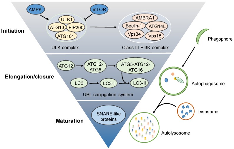
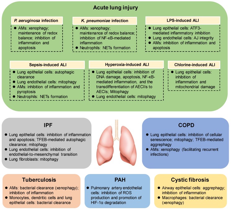
Autophagy is a highly conserved catabolic process involving autolysosomal degradation of cellular components, including protein aggregates, damaged organelles (such as mitochondria, endoplasmic reticulum, and others), as well as various pathogens. Thus, the autophagy pathway represents a major adaptive response for the maintenance of cellular and tissue homeostasis in response to numerous cellular stressors. A growing body of evidence suggests that autophagy is closely associated with diverse human diseases. Specifically, acute lung injury (ALI) and inflammatory responses caused by bacterial infection or xenobiotic inhalation (e.g., chlorine and cigarette smoke) have been reported to involve a spectrum of alterations in autophagy phenotypes. The role of autophagy in pulmonary infection and inflammatory diseases could be protective or harmful dependent on the conditions. In this review, we describe recent advances regarding the protective features of autophagy in pulmonary diseases, with a focus on ALI, idiopathic pulmonary fibrosis (IPF), chronic obstructive pulmonary disease (COPD), tuberculosis, pulmonary arterial hypertension (PAH) and cystic fibrosis.

Keywords: Autophagy, inflammation, acute lung injury, idiopathic pulmonary fibrosis, COPD, tuberculosis, PAH, cystic fibrosis.


Conflict of interest statement

The authors declare no conflict of interest.

Figures

Figure 1
Figure 1
Autophagy machinery. The autophagy process involves initiation, elongation/closure and maturation. The autophagy process is initiated by autophagosome biogenesis to form phagophore, which is regulated by the activation of the preinitiation complex (also known as the ULK complex, containing ULK1/2, ATG13, FIP200, and ATG101) and subsequent activation of the initiation complex (also called the class III PI3K complex, consisting of VPS34, VPS15, Beclin 1, ATG14L, and AMBRA1). The phagophore is then elongated and closed to form a double-membrane autophagosome, which is tightly regulated by the ubiquitin–like (UBL) conjugation systems. The autophagosome will fuse with a lysosome to form an autolysosome for degradation. The SNARE-like proteins may play important roles in autophagosome–lysosome degradation. AMPK—5′-AMP-activated protein kinase; mTOR—the mammalian target of rapamycin; ULK1—UNC51-like kinase 1; class III PI3K—the class III phosphatidylinositol-3-kinase.
Figure 2
Figure 2
The protective mechanisms of autophagy in lung diseases. Autophagy may provide a protective role in the pathogenesis of various lung diseases (including ALI, IPF, COPD, tuberculosis, PAH, cystic fibrosis, etc.), through regulating diverse biological events, including inflammatory response, redox balance, DNA damage repair, apoptosis, pyroptosis, cellular senescence, NETs formation, mitochondrial homeostasis, pathogen or aggresome clearance, etc. ALI—acute lung injury; IPF—idiopathic pulmonary fibrosis; COPD—chronic obstructive pulmonary disease; PAH—pulmonary arterial hypertension; TFEB—transcription factor EB; AMs—alveolar macrophages; NETs—neutrophil extracellular traps; AJ integrity—adherens junctional integrity; P. aeruginosaPseudomonas aeruginosa; K. pneumoniaeKlebsiella pneumoniae; LPS—lipopolysaccharide.

References

    1. Klionsky D.J., Abdelmohsen K., Abe A., Abedin M.J., Abeliovich H., Acevedo Arozena A., Adachi H., Adams C.M., Adams P.D., Adeli K., et al. Guidelines for the use and interpretation of assays for monitoring autophagy (3rdedition) Autophagy. 2016;12:1–222. doi: 10.1080/15548627.2015.1100356. - DOI - PMC - PubMed
    1. Gatica D., Lahiri V., Klionsky D.J. Cargo recognition and degradation by selective autophagy. Nat. Cell Biol. 2018;20:233–242. doi: 10.1038/s41556-018-0037-z. - DOI - PMC - PubMed
    1. Anding A.L., Baehrecke E.H. Cleaning House: Selective Autophagy of Organelles. Dev. Cell. 2017;41:10–22. doi: 10.1016/j.devcel.2017.02.016. - DOI - PMC - PubMed
    1. Green D.R., Levine B. To be or not to be? How selective autophagy and cell death govern cell fate. Cell. 2014;157:65–75. doi: 10.1016/j.cell.2014.02.049. - DOI - PMC - PubMed
    1. Morel E., Mehrpour M., Botti J., Dupont N., Hamai A., Nascimbeni A.C., Codogno P. Autophagy: A Druggable Process. Annu. Rev. Pharmacol. Toxicol. 2017;57:375–398. doi: 10.1146/annurev-pharmtox-010716-104936. - DOI - PubMed

Publication types

LinkOut - more resources