. 2019 Feb 4;38(1):52.
doi: 10.1186/s13046-019-1049-7.

CD36 mediates palmitate acid-induced metastasis of gastric cancer via AKT/GSK-3β/β-catenin pathway

Affiliations

CD36 mediates palmitate acid-induced metastasis of gastric cancer via AKT/GSK-3β/β-catenin pathway

Jiaomeng Pan et al. J Exp Clin Cancer Res. .

Abstract

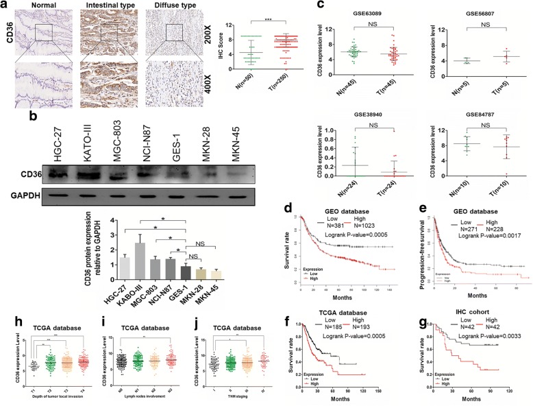
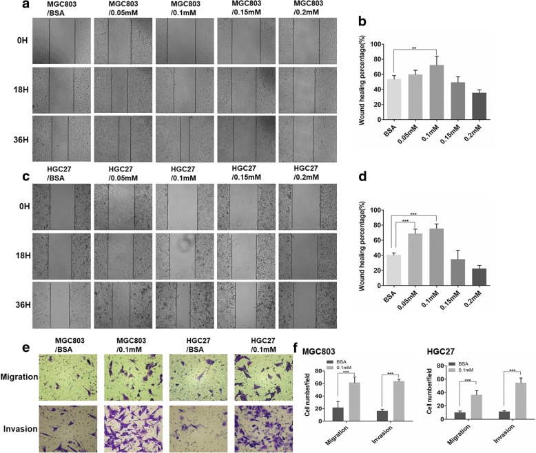
Background: Gastric cancer (GC) has a clear predilection for metastasis toward the omentum which is primarily composed of adipose tissue, indicating that fatty acids may contribute to this phenomenon. However their function remains poorly understood in GC. In this study, we investigated the role of palmitate acid (PA) and its cellular receptor CD36 in the progression of GC.

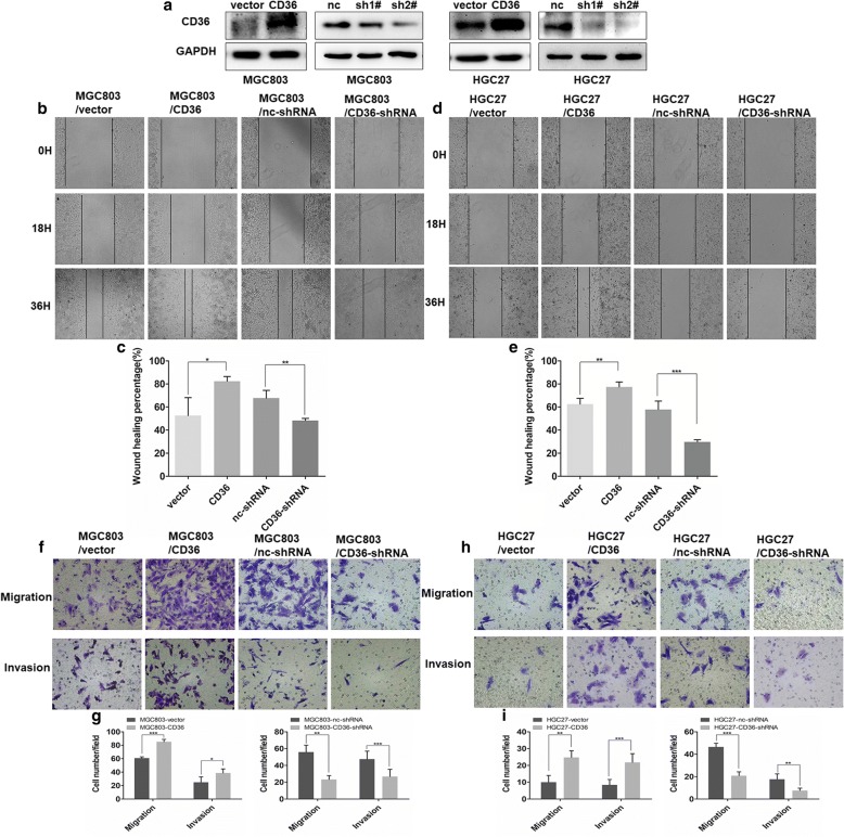
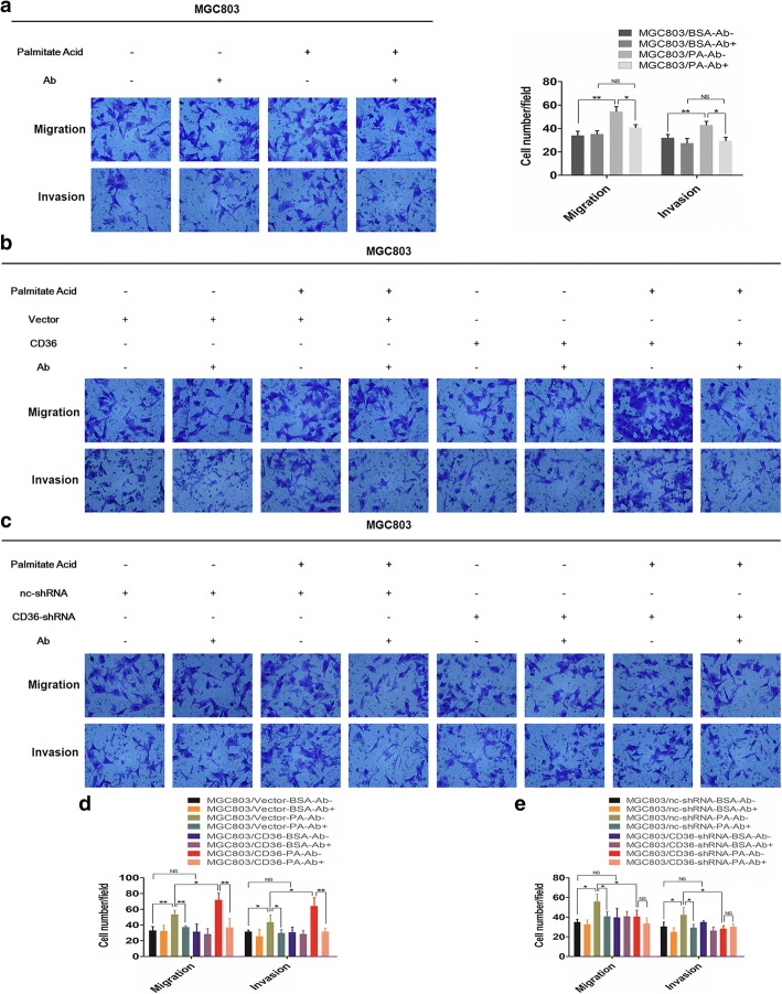
Methods: Immunohistochemical (IHC) staining was performed to detect CD36 expression in GC tissues and its clinical significance was determined statistically. CD36 over-expression and knock-down expression cell models were developed and tested in vitro. Wound-healing assays, migration assays, and invasion assays were performed and peritoneal implants into nude mice were done to assess the biological effects of PA and CD36. The underlying mechanisms were investigated using western blot, immunofluorescence (IF), quantitative real-time PCR (qRT-PCR) and antibody blocking assays.

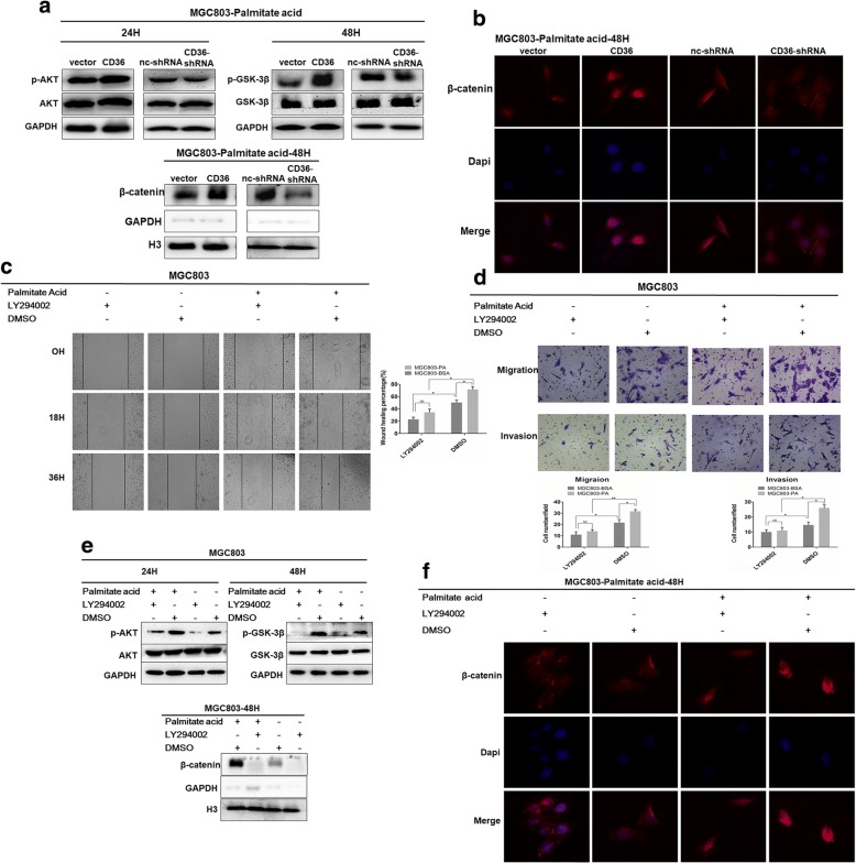
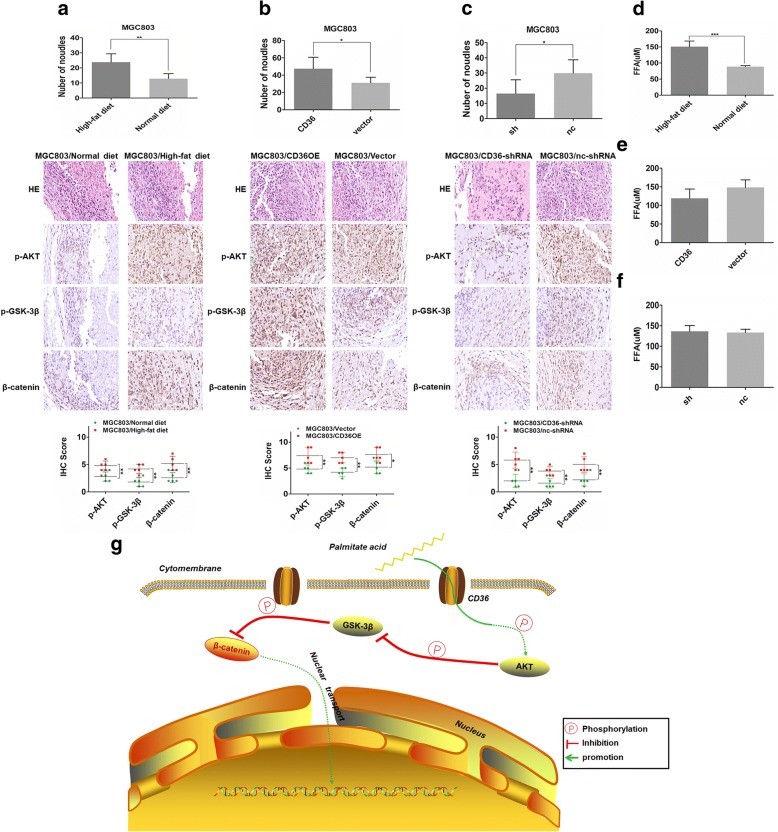
Results: PA promoted the metastasis of GC by phosphorylation of AKT, which facilitated the nuclear localization of β-catenin through inactivation of GSK-3β via phosphorylation. This tumor-promoting effect of PA was mediated by CD36, a cell surface receptor of fatty acids (FAs). The higher the CD36 expression levels in GC tissues correlated with the poorer the prognosis of patients according to the TCGA database, the GEO database and our own clinical data.

Conclusions: Our experiments established CD36 as a key mediator of FA-induced metastasis of GC via the AKT/GSK-3β/β-catenin signaling pathway. CD36 might, therefore, constitute a potential therapeutic target for clinical intervention in GC.

Keywords: AKT; CD36; GSK-3β; Gastric cancer; Metastasis; Palmitate acid; β-catenin.


Conflict of interest statement

Ethics approval

An institutional review board from the local Human Research Ethics Committee of Ruijin Hospital, Shanghai Jiao Tong University School of Medicine, China approved the study. All human participants provided informed consent. All animal experiments were also approved by the local Laboratory Animal Ethics Committee of Ruijin Hospital and conducted based on the Guide for the Care and Use Laboratory Animals of Ruijin Hospital. All samples tested for GC were anonymous in accordance with legal and ethical standards.

Consent for publication

All contributing authors agree to the publication of this article.

Competing interests

The authors declare that they have no competing interests.

Publisher’s Note

Springer Nature remains neutral with regard to jurisdictional claims in published maps and institutional affiliations.

Figures

Fig. 1
Fig. 1
Expression of CD36 in GC tissues, normal tissues and cell lines and its correlation with the clinico-pathology of GC. a IHC staining of CD36 in GC tissues and adjacent non-tumor tissues. Histogram shows the IHC scores of CD36 expression in GC tissues and normal tissues. b The expression level of CD36 in GC cell lines and immortalized gastric epithelial cell line (GES-1) by western blot and densitometric measurements of CD36 protein levels. c CD36 mRNA levels in GC tissues and paired normal tissues from GEO database (T = GC tissues, N = paired normal tissues). d, f and g Cumulative survival curves of GC patients from GEO database, TCGA database and our own clinical data. e PPS analysis of GC patients from GEO database. h, i and j Correlation between CD36 expression in tumor tissues and clinico-pathological parameters based on TCGA database. *P<0.05, **P<0.01, ***P<0.001, ‘NS’ means not significant
Fig. 2
Fig. 2
PA enhances the migratory and invasive abilities of GC cells. a and c Effect of different concentrations of PA on GC cell wound-healing (%) (mag. × 40). b and d Histograms of wound-healing (%) (mag. × 40) measured at 0, 18, and 36 h. e Effect of PA on GC cell migration and invasion (mag. × 200). f Histograms of the number of migrated and invaded cells (mag. × 200). Five random fields were selected for statistical analysis. Data are shown as mean ± SD of three independent experiments. *P<0.05, **P<0.01, ***P<0.001
Fig. 3
Fig. 3
CD36 mediates the migration and invasion of GC cells induced by PA. a Western blot analysis of CD36 expression in MGC803 and HGC27 cells transfected with CD36-plasmid, vector-plasmid control, nc-shRNA plasmid, CD36-shRNA#1 plasmid or CD36-shRNA#2 plasmid. b, d, f and h Effect of CD36 knockdown or overexpression on wound-healing of GC cells (mag. × 40), migration and invasion (mag. × 200). c, e, g and i Histograms of wound-healing (%) (mag. × 40) and the number of migrated and invaded cells (mag. × 200). Five random fields were selected for statistical analysis. Data are shown as mean ± SD of three independent experiments. *P<0.05, **P<0.01, ***P<0.001
Fig. 4
Fig. 4
CD36 promotes GC metastasis via uptake of PA. a Specific block of cellular FA uptake with anti-CD36 antibody inhibits PA-mediated migration and invasion of MGC803. Histogram shows the number of migrated and invaded cells (mag. × 200). Five random fields were selected for statistical analysis. b and c Block of cellular FA uptake by anti-CD36 inhibited migration and invasion of PA-treated GC cells (MGC803/Vector, MGC803/CD36, MGC803/nc-shRNA, MGC803/CD36-shRNA). d and e Histograms of the number of migrated and invaded cells (mag. × 200). Five random fields were selected for statistical analysis. Data are shown as mean ± SD, *P<0.05, **P<0.01, ***P<0.001, ‘NS’ means not significant
Fig. 5
Fig. 5
PA promotes GC cell migration and invasion via the AKT/GSK-3β/ β-catenin signaling pathway. a and c Effect of PA on protein levels of p-AKT, p-GSK-3β, AKT, GSK-3β and global β-catenin at different time points. b and d Densitometry showing effect of PA on protein levels of p-AKT, p-GSK-3β, AKT, GSK-3β and global β-catenin. e and f Effect of PA on cellular mRNA level of β-catenin at different time points. g and h Effects of PA on nuclear-transport of β-catenin at selected time points. Densitometry showing effect of PA on nuclear protein level of β-catenin. i and j IF showing effect of PA on cellular location of β-catenin in GC cells at selected time points. Data are shown as mean ± SD of three independent experiments. *P<0.05, **P<0.01, ***P<0.001, ‘NS’ means not significant
Fig. 6
Fig. 6
CD36 mediates PA-induced activation of AKT/GSK-3β/β-catenin signaling. a Effects of knockdown and overexpression of CD36 on p-AKT, AKT, p-GSK-3β, GSK-3β and nuclear β-catenin. b IF showing effects of knockdown and overexpression of CD36 on cellular location of β-catenin in PA-treated GC cells. c and d PI3K inhibitor, LY294002 inhibited migration and invasion of MGC803. Histograms show wound-healing (%) (mag. × 40) and number of migrated and invaded cells (mag. × 200). Five random fields were selected for statistical analysis. e Expression levels of p-AKT, AKT, p-GSK-3β, GSK-3β, nuclear β-catenin in PA-treated MGC803 in the presence of LY294002, compared to controls. f IF showing effects of LY294002 on cellular location of β-catenin in PA-treated GC cells, compared to controls. Data are shown as mean ± SD of three independent experiments. *P<0.05, **P<0.01, ***P<0.001, ‘NS’ means not significant
Fig. 7
Fig. 7
CD36 mediates PA-induced peritoneal dissemination in nude mice. a, b and c (upper) histograms of peritoneal tumor nodules four weeks after implantation of tumor cells (MGC803, MGC803/vector, MGC803/CD36, MGC803/nc-shRNA, MGC803/CD36-shRNA) into the peritoneum of nude mice. (middle) Histological appearance of disseminated tumors in the abdominal cavities of each group by H&E staining and IHC of p-AKT, p-GSK-3β, and β-catenin. (mag. × 400). (lower) Histograms show the IHC scores of p-AKT, p-GSK-3β and β-catenin in tumor tissues of each group. d, e and f Concentration of free long-chain FAs in the serum of mice. g CD36 mediates cellular uptake of PA which activates AKT through phosphorylation and then inactivates GSK-3β, which inhibits degradation of β-catenin and promotes its nuclear-transportation. Data are shown as mean ± SD. *P<0.05, **P<0.01, ***P<0.001

References

    1. den Hoed CM, Kuipers EJ. Gastric Cancer: how can we reduce the incidence of this disease? Curr Gastroenterol Rep. 2016;18:34. doi: 10.1007/s11894-016-0506-0. - DOI - PMC - PubMed
    1. Park JY, von Karsa L, Herrero R. Prevention strategies for gastric cancer: a global perspective. Clin Endosc. 2014;47:478–489. doi: 10.5946/ce.2014.47.6.478. - DOI - PMC - PubMed
    1. Torre LA, Bray F, Siegel RL, Ferlay J, Lortet-Tieulent J, Jemal A. Global cancer statistics, 2012. CA Cancer J Clin. 2015;65:87–108. doi: 10.3322/caac.21262. - DOI - PubMed
    1. Camarda R, Zhou AY, Kohnz RA, Balakrishnan S, Mahieu C, Anderton B, et al. Inhibition of fatty acid oxidation as a therapy for MYC-overexpressing triple-negative breast cancer. Nat Med. 2016;22:427–432. doi: 10.1038/nm.4055. - DOI - PMC - PubMed
    1. Hashimoto S, Mikami S, Sugino H, Yoshikawa A, Hashimoto A, Onodera Y, et al. Lysophosphatidic acid activates Arf6 to promote the mesenchymal malignancy of renal cancer. Nat Commun. 2016;7:10656. doi: 10.1038/ncomms10656. - DOI - PMC - PubMed

MeSH terms