

Hippocampal atrophy and intrinsic brain network dysfunction relate to alterations in mind wandering in neurodegeneration
- PMID: 30718430
- PMCID: PMC6386688
- DOI: 10.1073/pnas.1818523116
Hippocampal atrophy and intrinsic brain network dysfunction relate to alterations in mind wandering in neurodegeneration
Abstract
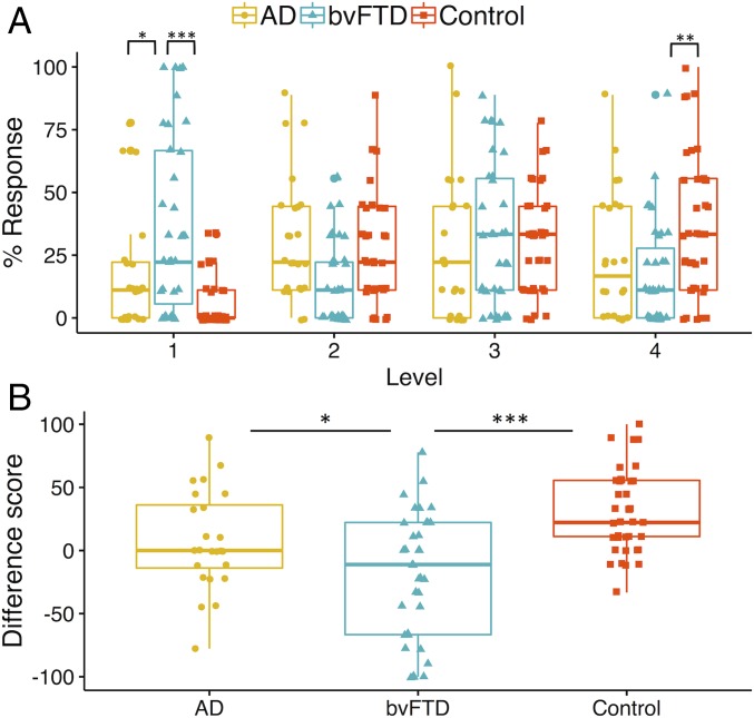
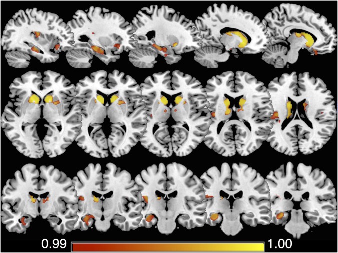
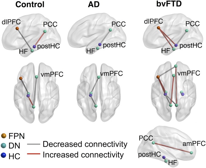
Mind wandering represents the human capacity for internally focused thought and relies upon the brain's default network and its interactions with attentional networks. Studies have characterized mind wandering in healthy people, yet there is limited understanding of how this capacity is affected in clinical populations. This paper used a validated thought-sampling task to probe mind wandering capacity in two neurodegenerative disorders: behavioral variant frontotemporal dementia [(bvFTD); n = 35] and Alzheimer's disease [(AD); n = 24], compared with older controls (n = 37). These patient groups were selected due to canonical structural and functional changes across sites of the default and frontoparietal networks and well-defined impairments in cognitive processes that support mind wandering. Relative to the controls, bvFTD patients displayed significantly reduced mind wandering capacity, offset by a significant increase in stimulus-bound thought. In contrast, AD patients demonstrated comparable levels of mind wandering to controls, in the context of a relatively subtle shift toward stimulus-/task-related forms of thought. In the patient groups, mind wandering was associated with gray matter integrity in the hippocampus/parahippocampus, striatum, insula, and orbitofrontal cortex. Resting-state functional connectivity revealed associations between mind wandering capacity and connectivity within and between regions of the frontoparietal and default networks with distinct patterns evident in patients vs. controls. These findings support a relationship between altered mind wandering capacity in neurodegenerative disorders and structural and functional integrity of the default and frontoparietal networks. This paper highlights a dimension of cognitive dysfunction not well documented in neurodegenerative disorders and validates current models of mind wandering in a clinical population.
Keywords: Alzheimer’s disease; behavioral variant frontotemporal dementia; default mode network; hippocampus; mind wandering.
Conflict of interest statement
The authors declare no conflict of interest.
Figures
References
-
- Kucyi A. Just a thought: How mind-wandering is represented in dynamic brain connectivity. Neuroimage. 2018;180:505–514. - PubMed
-
- Christoff K, Irving ZC, Fox KC, Spreng RN, Andrews-Hanna JR. Mind-wandering as spontaneous thought: A dynamic framework. Nat Rev Neurosci. 2016;17:718–731. - PubMed
-
- Fox KC, Spreng RN, Ellamil M, Andrews-Hanna JR, Christoff K. The wandering brain: Meta-analysis of functional neuroimaging studies of mind-wandering and related spontaneous thought processes. Neuroimage. 2015;111:611–621. - PubMed
Publication types
MeSH terms
Grants and funding
LinkOut - more resources
Full Text Sources
Medical
