. 2019 Feb 4;10(2):101.
doi: 10.1038/s41419-019-1343-5.

Generation and characterization of novel anti-DR4 and anti-DR5 antibodies developed by genetic immunization

Affiliations

Generation and characterization of novel anti-DR4 and anti-DR5 antibodies developed by genetic immunization

Agathe Dubuisson et al. Cell Death Dis. .

Abstract

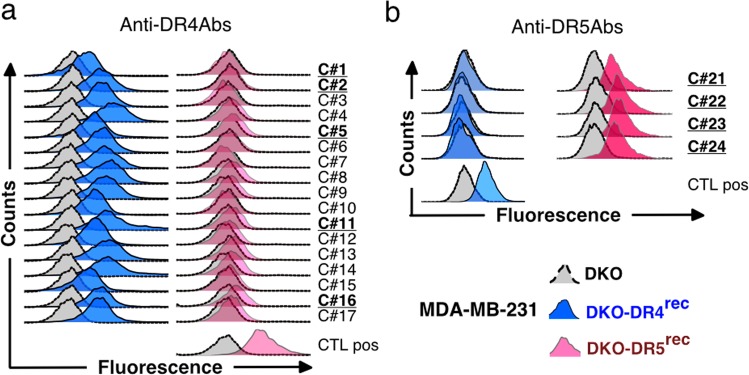
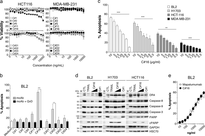
Development of therapeutic antibodies in oncology has attracted much interest in the past decades. More than 30 of them have been approved and are being used to treat patients suffering from cancer. Despite encouraging results, and albeit most clinical trials aiming at evaluating monoclonal antibodies directed against TRAIL agonist receptors have been discontinued, DR4 or DR5 remain interesting targets, since these receptors are overexpressed by tumour cells and are able to trigger their death. In an effort to develop novel and specific anti-DR4 and anti-DR5 antibodies with improved properties, we used genetic immunization to express native proteins in vivo. Injection of DR4 and DR5 cDNA into the tail veins of mice elicited significant humoral anti-DR4 and anti-DR5 responses and fusions of the corresponding spleens resulted in numerous hybridomas secreting antibodies that could specifically recognize DR4 or DR5 in their native forms. All antibodies bound specifically to their targets with a very high affinity, from picomolar to nanomolar range. Among the 21 anti-DR4 and anti-DR5 monoclonal antibodies that we have produced and purified, two displayed proapoptotic properties alone, five induced apoptosis after cross-linking, four were found to potentiate TRAIL-induced apoptosis and three displayed antiapoptotic potential. The most potent anti-DR4 antibody, C#16, was assessed in vivo and was found, alone, to inhibit tumour growth in animal models. This is the first demonstration that DNA-based immunization method can be used to generate novel monoclonal antibodies targeting receptors of the TNF superfamily that may constitute new therapeutic agents.


Conflict of interest statement

The authors declare no conflict of interest.

Figures

Fig. 1
Fig. 1. Cell surface receptor recognition of the produced anti-DR4 and anti-DR5 antibodies.
MDA-MB-231 DKO (grey curve) and MDA-MB-231 DKO reconstituted with either DR4 (MDA-MB-231-DKO-DR4rec, blue curve) or DR5 (MDA-MB-231-DKO-DR5rec, red curve) were stained with a anti-DR4 antibodies or b anti-DR5 antibodies. Primary antibodies were then detected using a AF488-conjugated mouse-specific IgG (H+L) secondary antibody and analysed by flow cytometry
Fig. 2
Fig. 2. Apoptosis induced by anti-DR4 or -DR5 antibodies.
a Viability of HCT-116 and MDA-MB-231 were determined using methylene blue, 16 h after treatment with increasing concentrations of anti-DR4 mAbs (C#1, C#2, C#5, C#11 and C#16) or anti-DR5 mAbs (C#21, C#22, C#23 and C#24). Values are means ± SD (n = 3). b BL2 cells were pre-incubated or not for 30 min at 37 °C with 10 µM of Q-VD-Oph prior stimulation for 20 h with 10 µg/mL of mAbs. Cells were next stained with Annexin V/7AAD and apoptosis was quantified by flow cytometry. c BL2, H1703, HCT116 and MDA-MB-231 cell lines were treated for 20 h with increasing concentrations of C#16, and apoptosis was quantified as above by flow cytometry. d BL2, H1703 and HCT116 cells were stimulated with 1 µg/mL His-TRAIL, 1 or 5 µg/mL of C#16 or 5 µg/mL C#1 for 8 h. Corresponding cell extracts were analysed by immunoblot. e BL2 cells were treated with increasing concentration of C#16 or Mapatumumab for 20 h, and apoptosis was quantified with Annexin V/7AAD staining by flow cytometry. All values are presented here as ±SD (n = 3). Significance was evaluated by analysis of variance tests. *p < 0.1, **p < 0.05, ***p < 0.01
Fig. 3
Fig. 3. The anti-DR4 antibody C#16 reduces tumour growth in xenograft models.
BL2 cells mixed with Matrigel were inoculated into the right flank of mice subcutaneously at day 0. Mice were then randomized into groups of five to six mice and were injected intraperitoneally with C#16 (10 mg/kg) or vehicle control (phosphate-buffered saline) at the indicated times (red arrows). Each time point represents the mean value (±s.e.m) of the tumour sizes on the day of measurement. The animals were sacrificed 34 days postinoculation. a Pre-established xenograft tumour growth and corresponding mouse body weight. Significance was evaluated by analysis of variance (ANOVA) test. **p < 0.05. b De novo model tumour growth and corresponding mouse body weight. Significance was evaluated by ANOVA test. ****p < 0.0001. c Comparison of tumour weights from de novo model between mice injected with saline and mice injected with C#16 (10 mg/kg). ***p < 0.01. Significance was tested using unpaired t test
Fig. 4
Fig. 4. Anti-DR4 or anti-DR5 antibodies can potentiate or inhibit tumour necrosis factor-related apoptosis inducing ligand (TRAIL) action.
Cell viability of a HCT-116 and b MDA-MB-231 cells was determined using methylene blue test. Cells were treated for 16 h with the indicated concentration of DR4 (left) or DR5 (right) monoclonal antibodies (mAbs) in combination with His-TRAIL. Values are means ± SD (n = 3). c Left: BL2 cells were treated in 24-well plates for 20 h with 1 µg/mL of soluble TRAIL or with a combination of 10 µg/mL of mAbs and 1 µg/mL His-TRAIL. When indicated, 10 µM of Q-VD-Oph was added prior treatment for 30 min at 37 °C. Medium and cells were harvested and apoptosis was detected by Annexin V/7AAD staining and flow cytometry. Significance was evaluated by analysis of variance (ANOVA) test with the mean values ± SD (n = 3) **p < 0.05, ***p < 0.01. Right: BL2 were stimulated for 8 h with 5 µg/mL of C#2, C#11, C#22 or C#24 in the presence or absence of 1 µg/mL His-TRAIL and cleavage of effector caspase-3 and PARP-1 was analysed by immunoblot. GAPDH was used as a gel-loading control. d MDA-MB-231 cells were treated for 8 h with 5 µg/mL C#2, C#11, C#22 or C#24 in the presence or absence of 1 µg/mL His-TRAIL and apoptosis (upper part) or caspase activation (lower part) were analysed as above by flow cytometry or immunoblot. e HCT116-DKO cells reconstituted with only DR5 (HCT116 DKO-DR5rec) were treated with 10 µg/mL of C#21 or C#23 in the presence or absence of 1 µg/mL His-TRAIL and apoptosis was quantified by flow cytometry. Significance was evaluated by ANOVA test with the mean values ± SD (n = 3) *p < 0.1, **p < 0.05
Fig. 5
Fig. 5. Affinity evaluation of monoclonal antibodies (mAbs).
a The binding profile of mAbs towards DR4-Fc and DR5-Fc was determined by biolayer interferometry assay. The coloured lines represent the binding response signal at various mAb concentrations (from 500 to 7.8 nM) against DR4-Fc or DR5-Fc. Binding of mAbs was monitored in real time to obtain on (Kon) and off (Koff) rates. Association was measured for 120 s while dissociation was measured for 600 s. The equilibrium constant (KD) was calculated as Koff/Kon using 1:1 binding stoichiometric model. b On–off rate map indicating the binding kinetic parameters of anti-DR4 and anti-DR5 antibodies. The association rate constant (Kon) is plotted against the dissociation rate constant (Koff). The diagonal lines indicate the equilibrium dissociation constant (KD). Agonists antibodies (C#16 and C#22) are shown in orange and enhancer antibodies (C#2, C#11, C#22 and C#24) are shown in green

References

    1. French LE, Tschopp J. The TRAIL to selective tumor death. Nat. Med. 1999;5:146–147. doi: 10.1038/5505. - DOI - PubMed
    1. Walczak H, et al. Tumoricidal activity of tumor necrosis factor-related apoptosis-inducing ligand in vivo. Nat. Med. 1999;5:157–163. doi: 10.1038/5517. - DOI - PubMed
    1. Micheau O, Shirley S, Dufour F. Death receptors as targets in cancer. Br. J. Pharmacol. 2013;169:1723–1744. doi: 10.1111/bph.12238. - DOI - PMC - PubMed
    1. Dubuisson, A. & Micheau, O. Antibodies and derivatives targeting DR4 and DR5 for cancer therapy. Antibodies6, 16 (2017). - PMC - PubMed
    1. Leng Y, et al. Circularly permuted TRAIL plus thalidomide and dexamethasone versus thalidomide and dexamethasone for relapsed/refractory multiple myeloma: a phase 2 study. Cancer Chemother. Pharmacol. 2017;79:1141–1149. doi: 10.1007/s00280-017-3310-0. - DOI - PubMed

Publication types

MeSH terms

Substances