. 2019 Feb 4;9(1):1121.
doi: 10.1038/s41598-018-37439-0.

A cancer-associated point mutation disables the steric gate of human PrimPol

Affiliations

A cancer-associated point mutation disables the steric gate of human PrimPol

Alberto Díaz-Talavera et al. Sci Rep. .

Abstract

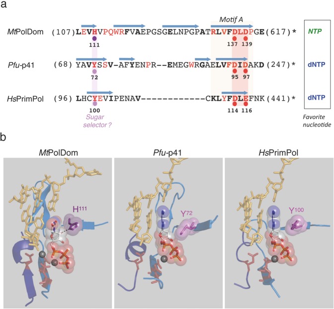
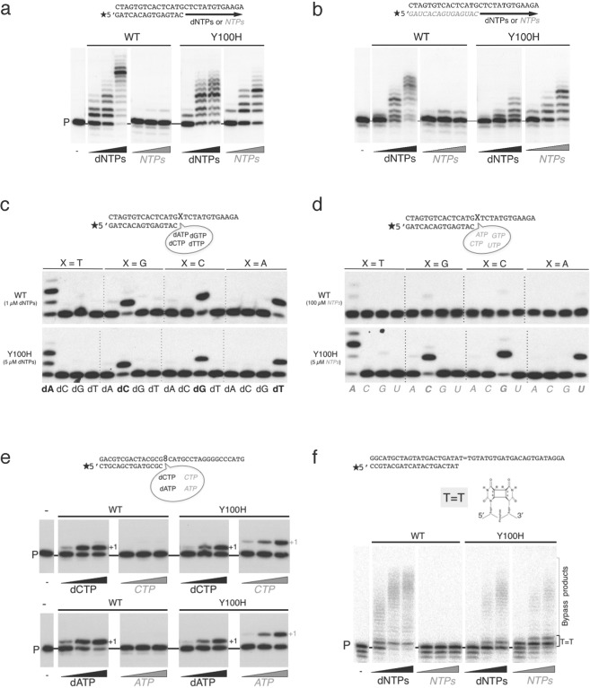
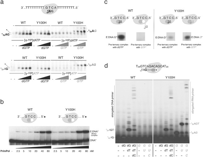
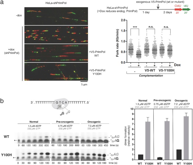
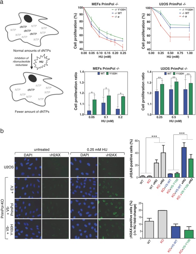
PrimPol is a human primase/polymerase specialized in re-starting stalled forks by repriming beyond lesions such as pyrimidine dimers, and replication-perturbing structures including G-quadruplexes and R-loops. Unlike most conventional primases, PrimPol proficiently discriminates against ribonucleotides (NTPs), being able to start synthesis using deoxynucleotides (dNTPs), yet the structural basis and physiological implications for this discrimination are not understood. In silico analyses based on the three-dimensional structure of human PrimPol and related enzymes enabled us to predict a single residue, Tyr100, as the main effector of sugar discrimination in human PrimPol and a change of Tyr100 to histidine to boost the efficiency of NTP incorporation. We show here that the Y100H mutation profoundly stimulates NTP incorporation by human PrimPol, with an efficiency similar to that for dNTP incorporation during both primase and polymerase reactions in vitro. As expected from the higher cellular concentration of NTPs relative to dNTPs, Y100H expression in mouse embryonic fibroblasts and U2OS osteosarcoma cells caused enhanced resistance to hydroxyurea, which decreases the dNTP pool levels in S-phase. Remarkably, the Y100H PrimPol mutation has been identified in cancer, suggesting that this mutation could be selected to promote survival at early stages of tumorigenesis, which is characterized by depleted dNTP pools.


Conflict of interest statement

The authors declare no competing interests.

Figures

Figure 1
Figure 1
Prediction of the sugar selector in human PrimPol. (a) Primary sequence comparison of the region encompassing motif A of MtPolDom, Pfu-p41 and HsPrimPol. Catalytic residues involved in metal binding are indicated with red dots. Single residues acting as a sugar selector favoring NTPs or dNTPs are indicated with violet or pink dots, respectively; β-strands are indicated as light blue arrows. Figures in parenthesis indicate the number of N-terminal or C-terminal amino acid residues that are not shown. Invariant (red) or conserved (bold black) residues are indicated (see also Supplemental Fig. 1). (b) Structural details of the region aligned in part a, containing candidate residues to act as sugar selectors, and two catalytic metal ligands; a third metal ligand, embedded in an additional peptide segment (motif C; depicted in dark blue) is also shown in MtPolDom (PDB ID: 3PKY, DNA template/primer from 4MKY and NTP from 3PKY), Pfu-p41 (PDB ID: 1G71, DNA template/primer and dNTP from 5L2X) and HsPrimPol (PDB ID: 5L2X, DNA template/primer and dNTP from 5L2X).
Figure 2
Figure 2
Use of ribonucleotides by the Y100H variant during primer extension and translesion synthesis. (a) DNA primer extension assay on the indicated template/primer by either wild-type (WT) PrimPol or Y100H, using increasing concentration of dNTPs (1, 10, 50 µM) or NTPs (1, 10, 50 µM). (b) RNA primer extension on the indicated template/ RNA primer structure as described in part a. (c) Matched versus mismatched nucleotide insertion at the four template bases (indicated as X in the scheme). Nucleotide insertion on each template-primer was analyzed in the presence of each individual dNTP at 1 µM or 5 µM for the WT or Y100H mutant respectively. (d) As described in (c), using NTP as substrates at 100 µM or 5 µM for the WT or Y100H mutant respectively. (e) Lesion bypass of 8oxoG (scheme of the template/primer structure at the top) by either WT PrimPol or Y100H (100 nM) in the presence of 100 µM MnCl2 as metal cofactor with increasing concentrations (1, 10, 50 µM) of dCTP or CTP (upper panel) and dATP or ATP (lower panel). (f) Lesion bypass of a CPD lesion (scheme of the template/primer structure at the top) of either WT PrimPol or Y100H with increasing concentrations of dNTPs or NTPs (1, 10, 100 µM). Full length gels corresponding to parts a, b, e and f are shown in Supplemental Fig. 5. The autoradiographs shown in this figure are representative of at least 3 independent experiments.
Figure 3
Figure 3
Ribonucleotides are valid substrates for the Y100H variant during primer synthesis. (a) Scheme on the top shows PrimPol in complex with the GTCA template oligonucleotide and the two nucleotides forming the initial dimer. The autoradiograph shows dimer formation (primase activity) either by wild-type (WT) PrimPol or Y100H (400 nM) using [α-32P]dATP (upper panel) or [γ-32P]ATP (lower panel) as the 5′-site nucleotide (16 nM), and increasing concentrations of either dGTP or GTP as the incoming 3′-site nucleotide (0, 10, 50, 100 µM). (b) Binary complex formation, measured by EMSA, between WT PrimPol or Y100H and labeled 60-mer DNA template GTCC (1 nM), using the indicated PrimPol concentration (2.5, 5, 10, 20, 40 and 80 nM) (c) Pre-ternary complex formation measured by EMSA between WT PrimPol or Y100H (1 µM), 60-mer DNA template GTCC and either [α-32P]dGTP or [α-32P]GTP (16 nM). (d) DNA or RNA primers synthesized using as template 5′-T20ACGACAGACTGT29 -3′ to allow elongation beyond the dimer. Products were labeled with [γ-32P]ATP (16 nM) as the 5′nucleotide and each subsequent nucleotide (dGTP, dTTP, dCTP) was added (10 µM) as indicated in the figure. Full length gels corresponding to parts a to d are shown in Supplemental Fig. 6. The autoradiographs shown in this figure are representative of at least 3 independent experiments.
Figure 4
Figure 4
Y100H variant is competent for re-priming in vivo. (a) Workflow of the experimental design to measure replication fork rate by DNA fiber analysis after downregulation of endogenous PrimPol and expression of exogenous wild-type (WT) PrimPol or Y100H mutant. Fork rate values were calculated from the green length of red-green tracks. N > 300 values in each condition; n.s.: non-significant; ***p < 0.001 (Mann-Whitney test). Representative images of the different conditions are shown. (b) Dimer formation (primase assay) carried out using a 29-mer DNA template GTCA by either WT PrimPol or Y100H, in the presence of [γ-32P]ATP (16 nM) as the 5′nucleotide, and different physiological concentrations of either dGTP and GTP, as the incoming 3′nucleotide (normal cell concentration: 1.5 µM dGTP + 232 µM GTP, pre-oncogenic cell: 0.75 µM dGTP + 232 µM GTP or oncogenic cell: 7.2 µM dGTP + 232 µM GTP). The histogram shows the relative velocity of total dimer formation (AdG and AG) in the primase assays shown at the left. Full length gels corresponding to b are shown in Supplemental Fig. 7. The autoradiographs shown in this figure are representative of at least 3 independent experiments.
Figure 5
Figure 5
PrimPol Y100H enhances cellular resistance to dNTP pool depletion by reducing DSBs. (a) Left panels: schematic representation of the effect of hydroxyurea (HU) in altering the dNTPs/NTPs ratio. Central/upper panel: relative cell proliferation curves of PrimPol−/− MEFs transfected with empty vector (red), WT PrimPol (blue) or Y100H mutant (green), in the presence of increasing concentrations of HU (0.05, 0.10, 0.15, 0.20 and 0.25 mM); histogram in the central/lower panel shows the ratio of cell proliferation relative to WT PrimPol−/− cells at three HU concentrations (0.05, 0.1 and 0.2 mM). Right upper panel: relative cell proliferation curves of PrimPol−/− U2OS cells transfected as described above, in the presence of increasing concentrations of HU (0.25, 0.5 and 1 mM); the histogram in the right lower panel shows the cell proliferation ratio relative to WT PrimPol−/− cells at the same HU concentrations. t test **p < 0.01. (b) Left: representative confocal microscopy images of DAPI and γH2AX stainings in U2OS cells or PrimPol KO cells transfected with empty vector, WT PrimPol or Y100H mutant. When indicated, cells were incubated with 0.25 mM HU. Right: top histogram indicates the average median value of γH2AX intensity in each condition, derived from three biological replicates (>100 cells scored per condition in each replicate). Statistical significance was assessed with ANOVA and Bonferroni post-test. All pair-wise differences between lanes 1–2 and 3–4, or between 5–6 and 7–8, were significative. Bottom histogram depicts the fold-change difference in the intensity of γH2AX staining in the presence or absence of HU in each case.

References

    1. Clausen AR, Murray MS, Passer AR, Pedersen LC, Kunkel TA. Structure–function analysis of ribonucleotide bypass by B family DNA replicases. Proc Natl Acad Sci USA. 2013;110:16802–16807. doi: 10.1073/pnas.1309119110. - DOI - PMC - PubMed
    1. Nick McElhinny SA, et al. Abundant ribonucleotide incorporation into DNA by yeast replicative polymerases. Proc Natl Acad Sci USA. 2010;107:4949–4954. doi: 10.1073/pnas.0914857107. - DOI - PMC - PubMed
    1. Watt DL, Johansson E, Burgers PM, Kunkel TA. Replication of ribonucleotide-containing DNA templates by yeast replicative polymerases. DNA Repair (Amst) 2011;10:897–902. doi: 10.1016/j.dnarep.2011.05.009. - DOI - PMC - PubMed
    1. Hovatter KR, Martinson HG. Ribonucleotide-induced helical alteration in DNA prevents nucleosome formation. Proc Natl Acad Sci USA. 1987;84:1162–1166. doi: 10.1073/pnas.84.5.1162. - DOI - PMC - PubMed
    1. Li Y, Breaker RR. Kinetics of RNA degradation by specific base catalysis of transesterification involving the 2′-hydroxyl group. J Am Chem Soc. 1999;121:5364–5372. doi: 10.1021/ja990592p. - DOI

Publication types

MeSH terms