. 2019 Feb 4;9(1):1163.
doi: 10.1038/s41598-018-37473-y.

Mycobacterium tuberculosis Rv0366c-Rv0367c encodes a non-canonical PezAT-like toxin-antitoxin pair

Affiliations

Mycobacterium tuberculosis Rv0366c-Rv0367c encodes a non-canonical PezAT-like toxin-antitoxin pair

Himani Tandon et al. Sci Rep. .

Abstract

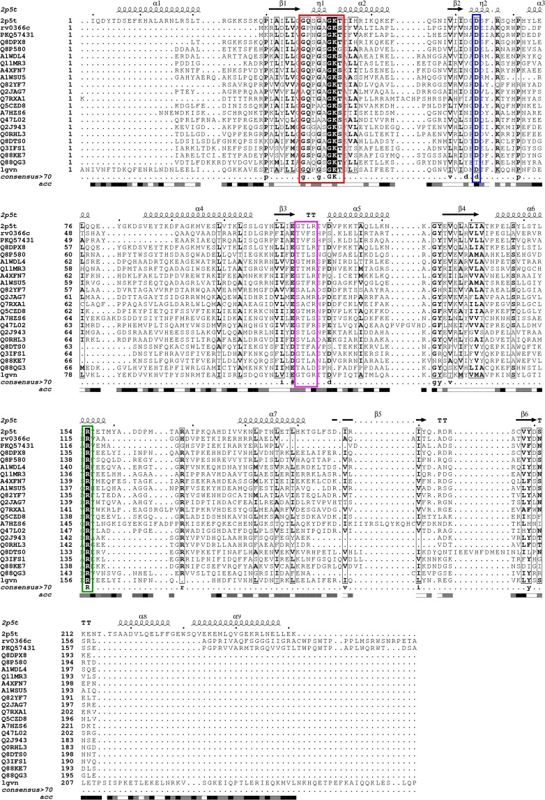
Toxin-antitoxin (TA) systems are ubiquitously existing addiction modules with essential roles in bacterial persistence and virulence. The genome of Mycobacterium tuberculosis encodes approximately 79 TA systems. Through computational and experimental investigations, we report for the first time that Rv0366c-Rv0367c is a non-canonical PezAT-like toxin-antitoxin system in M. tuberculosis. Homology searches with known PezT homologues revealed that residues implicated in nucleotide, antitoxin-binding and catalysis are conserved in Rv0366c. Unlike canonical PezA antitoxins, the N-terminal of Rv0367c is predicted to adopt the ribbon-helix-helix (RHH) motif for deoxyribonucleic acid (DNA) recognition. Further, the modelled complex predicts that the interactions between PezT and PezA involve conserved residues. We performed a large-scale search in sequences encoded in 101 mycobacterial and 4500 prokaryotic genomes and show that such an atypical PezAT organization is conserved in 20 other mycobacterial organisms and in families of class Actinobacteria. We also demonstrate that overexpression of Rv0366c induces bacteriostasis and this growth defect could be restored upon co-expression of cognate antitoxin, Rv0367c. Further, we also observed that inducible expression of Rv0366c in Mycobacterium smegmatis results in decreased cell-length and enhanced tolerance against a front-line tuberculosis (TB) drug, ethambutol. Taken together, we have identified and functionally characterized a novel non-canonical TA system from M. tuberculosis.


Conflict of interest statement

The authors declare no competing interests.

Figures

Figure 1
Figure 1
Representation of genomic region, ORF and promoters. The genomic locus for Rv0368c, Rv0367c, Rv0366c and Rv0365c are shown. Rv0367c, Rv0366c and Rv0365c were predicted to lie in the same operon. The promoter region was predicted to lie 26 bp upstream of Rv0367c. The annotation for these gene neighbours, obtained from Tuberculist is also given below the figure. Rv0367c was considered as a putative antitoxin for further analysis.
Figure 2
Figure 2
Structure-guided alignment of Rv0366c with ζ toxin from Streptococcus pyogenes, PezT from Streptococcus pneumoniae and other homologues. First sequence is the sequence of PezT of known structure. The nucleotide-binding motif is completely conserved and marked in red. The aspartate residue, important for deprotonation of substrate, is also conserved and marked in blue. The conserved arginine residue, marked in green, is important for both antitoxin-binding and catalysis. The other UNAG-binding residues are marked in pink. Other fully conserved residues are shown in black background, while semi-conserved residues in black boxes. Alignment was generated using ESpript 3.0.
Figure 3
Figure 3
Modelled structure for PezTMtb and comparison of substrate-binding pockets of known ζ toxins with PezTMtb. (A) Putative nucleotide-binding residues (black), conserved residue that may deprotonate the substrate (blue), residue with a potential role in both antitoxin-binding and catalysis (green) as well as solvent exposed, conserved residues, which include Lys40, Trp43, Pro44, His122, Val77 and Asn34 (yellow) are highlighted in the modelled PezTMtb structure. (B) Ribbon representation of superposed crystal structures of PezT (2p5t), ζ_ng (6epg), AvrRxo1 (4z8v) and the modelled structure of PezTMtb. The nucleotide-binding P-loop motif is shown in cartoon representation, cyan- PezT, blue – ζ_ng, yellow- AvrRxo1 and red – PezTMtb. The substrate-binding residues are shown in ball and stick, magenta- PezT, grey – ζ_ng, green - AvrRxo1 and orange– PezTMtb. The conserved Asp residue, important for substrate-binding/deprotonating the substrate, is shown as sticks in light-blue. Other two residues (Lys40 and Arg116) known to bind substrate and are conserved in PezTMtb are also labelled.
Figure 4
Figure 4
Alignment of PezAMtb with ε and PezA from other organisms. (A) The secondary structures predicted by PSIPRED show a ribbon-helix-helix (RHH) for the N-terminal region of Rv0367c. C-terminal region is also predicted to be ordered with α-helices and strands. (B) shows the multiple sequence alignment of C-terminal region of Rv0367c with ε and its homologues, and the C-terminal of PezA. Structural alignment of both ε and PezA were used to guide the alignment. Putative toxin-binding residue is marked in green and the residue predicted to occlude the nucleotide binding site is shown in blue. Other fully conserved residues are shown in black background, while semi-conserved residues in black boxes. Alignment was generated using ESpript 3.0.
Figure 5
Figure 5
Alignment of PezAMtb with PezA homologues (A) and ω from S. pyogenes (B). (A) Shows alignment of Rv0367c (O53702) with full-length PezA and its homologues. Though the N-terminal region shows sequence conservation, many residues that are a part of the helix-turn-helix motif in the N-terminal of PezA are not conserved in PezAMtb. (B) Shows alignment of PezAMtb (N-terminal) with ω repressor (1rq) of ε/ ζ TA system. Crystal structure of ω was used to guide the alignment. The fully conserved residues between these sequences are highlighted in black. Alignment was generated using ESpript 3.0.
Figure 6
Figure 6
Effect of overexpression of PezTMtb on bacterial growth. (A) E. coli BL-21, plysS, λDE3 was transformed with either pET28b or pET28b-pezTMtb. The expression of PezTMtb was induced by addition of 1 mM IPTG and growth was monitored by measuring OD600nm at indicated time points. (B,C) Expression of PezTMtb in both M. smegmatis mc2155 and M. tuberculosis H37Rv was induced by addition of 50 ng/ml anhydrotetracycline in liquid cultures. The growth of induced cultures was monitored by measuring OD600nm. (D,E) For co-expression studies, the expression of PezTMtb and PezAMtb was induced by addition of 50 ng/ml anhydrotetracycline and 0.2% acetamide, respectively. The growth of induced cultures was monitored by measuring OD600nm (D) and viable counts (E). For bacterial enumeration 10.0-fold serial dilutions were prepared and plated on MB7H11 plates. The plates were incubated at 37 °C for 2–3 days. The data shown in this panel is representative of two independent experiments. (F) M. smegmatis strains harbouring vector or PezTMtb or PezTMtbK15A or PezTMtbD36A or PezTMtbR116A were grown in MB7H9 medium till OD600nm of 0.2. The effect of expression of wild type and mutant PezTMtb proteins on M. smegmatis growth was monitored by measuring OD600nm. The data shown in these panels is representative of three independent experiments.
Figure 7
Figure 7
Effect of PezTMtb overexpression on cell length and drug tolerance of M. smegmatis. (A) DAPI stained fixed images of M. smegmatis harbouring vector or expressing PezTMtb in the absence or presence of PezAMtb after 3 hrs of induction. (B,C) The panel depicts cell length of M. smegmatis harbouring either pTetR-Int or pTetR-Int-PezTMtb or pTetR-Int-PezTMtb and pLam12-PezAMtb. The cultures were induced for either 3 hrs (B) or 6 hrs (C) and bacilli length was measured using Fluoview FV1000 software. The y-axis represents mean ± S.E. of cell length for bacilli per strain. The data shown in this panel is representative of three independent experiments. (D,E) Induced M. smegmatis cultures harbouring either pTetR-Int or pTetR-Int-PezTMtb were exposed to either 15.6 μM ethambutol or 2.0 μM levofloxacin. For bacterial enumeration, 12 hrs post-exposure, cultures were harvested, washed and 100 μl of 10.0-fold serial dilutions were plated on MB7H11 plates at 37 °C for 2–3 days. Significant differences were observed for the indicated groups (paired, two-tailed, t-test, *P < 0.05, ***P < 0.001).
Figure 8
Figure 8
Distribution of PezTMtb (upper panel) and PezAMtb (lower panel) in the genus Mycobacterium. The hits were obtained at an e-value better than 1e−04 and query coverage better than 70% from TBLASTN. The organisms belonging to MTBC are marked in green in both panels. Red marking indicates the organisms in which maximum number of other H37Rv toxin-antitoxin systems have been found to be conserved (unpublished). The other colours represent the habitat that these organisms belong to: cyan – water, brown – soil, pink – plants, black – air.

References

    1. Sala A, Bordes P, Genevaux P. Multiple toxin-antitoxin systems in Mycobacterium tuberculosis. Toxins (Basel) 2014;6:1002–1020. doi: 10.3390/toxins6031002. - DOI - PMC - PubMed
    1. Gerdes K, Christensen SK, Løbner-Olesen A. Prokaryotic toxin-antitoxin stress response loci. Nature Reviews Microbiology. 2005;3:371–382. doi: 10.1038/nrmicro1147. - DOI - PubMed
    1. Blower TR, Salmond GPC, Luisi BF. Balancing at survival’s edge: The structure and adaptive benefits of prokaryotic toxin-antitoxin partners. Curr. Opin. Struct. Biol. 2011;21:109–118. doi: 10.1016/j.sbi.2010.10.009. - DOI - PubMed
    1. Harms, A., Maisonneuve, E. & Gerdes, K. Mechanisms of bacterial persistence during stress and antibiotic exposure. Science (80-.). 354 (2016). - PubMed
    1. Melderen, L. V. et al. Molecular Basis of Gyrase Poisoning by the Addiction Toxin CcdB. 1091–1102, 10.1016/j.jmb.2005.03.049 (2005). - PubMed

Publication types

MeSH terms

Substances