. 2019 Feb 4;9(1):1220.
doi: 10.1038/s41598-018-37409-6.

Endogenous Neurosteroid (3α,5α)3-Hydroxypregnan-20-one Inhibits Toll-like-4 Receptor Activation and Pro-inflammatory Signaling in Macrophages and Brain

Affiliations

Endogenous Neurosteroid (3α,5α)3-Hydroxypregnan-20-one Inhibits Toll-like-4 Receptor Activation and Pro-inflammatory Signaling in Macrophages and Brain

Irina Balan et al. Sci Rep. .

Abstract

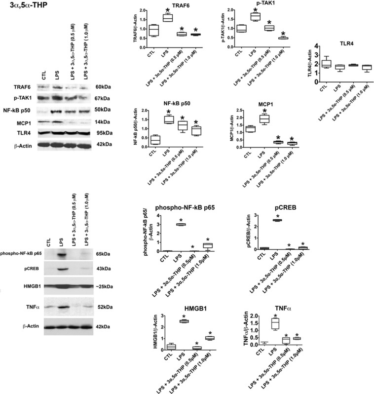
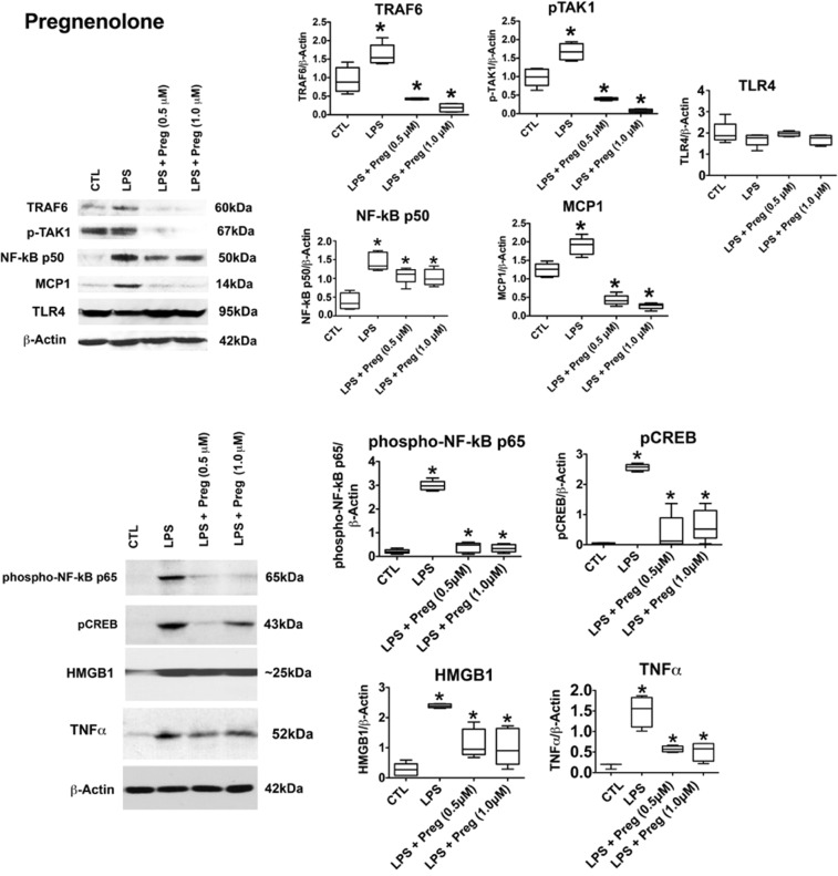
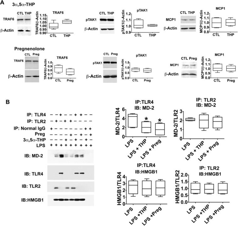
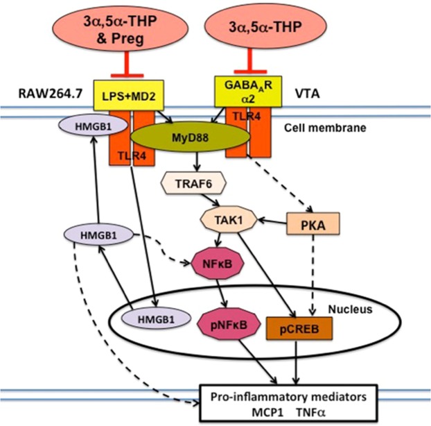
The endogenous neurosteroid (3α,5α)3-hydroxypregnan-20-one (3α,5α-THP, allopregnanolone) has protective activity in animal models of alcoholism, depression, traumatic brain injury, schizophrenia, multiple sclerosis, and Alzheimer's disease that is poorly understood. Because these conditions involve proinflammatory signaling through toll-like receptors (TLRs), we examined the effects of 3α,5α-THP, and pregnenolone on TLR4 activation in both the periphery and the central nervous system (CNS). We used monocytes/macrophages (RAW264.7) as a model of peripheral immune signaling and studied innately activated TLR4 in the ventral tegmental area (VTA) of selectively bred alcohol-preferring (P) rats. LPS activated the TLR4 pathway in RAW264.7 cells as evidenced by increased levels of p-TAK1, TRAF6, NF-κB p50, phospho-NF-κB- p65, pCREB, HMGB1, and inflammatory mediators, including MCP-1 and TNFα. Both 3α,5α-THP and pregnenolone (0.5-1.0μM) substantially (~80%) inhibited these effects, indicating pronounced inhibition of TLR4 signaling. The mechanism of inhibition appears to involve blockade of TLR4/MD-2 protein interactions in RAW246.7 cells. In VTA, 3α,5α-THP (15 mg/kg, IP) administration reduced TRAF6 (~20%), CRF (~30%), and MCP-1 (~20%) levels, as well as TLR4 binding to GABAA receptor α2 subunits (~60%) and MyD88 (~40%). The data suggest that inhibition of proinflammatory neuroimmune signaling underlies protective effects of 3α,5α-THP in immune cells and brain, apparently involving blocking of protein-protein interactions that initiate TLR4-dependent signaling. Inhibition of pro-inflammatory TLR4 activation represents a new mechanism of 3α,5α-THP action in the periphery and the brain.


Conflict of interest statement

The authors have filed for patent protection of the data in this report (Provisional patent #62/674,379). The authors declare no other conflicts of interest.

Figures

Figure 1
Figure 1
3α,5α-THP inhibits LPS-activated TLR4 signaling in RAW264.7 cells. 3α,5α-THP inhibits LPS-activated TLR4 signaling in RAW264.7 cells. RAW264.7 cells were treated with LPS alone (1 µg/ml) or LPS together with 3α,5α-THP (0.5 µM or 1 µM) and harvested after 24 hrs. The levels of p-TAK1 [F19 = 50.47, n = 5/grp], MCP-1 [F19 = 97.27, n = 5/grp], TRAF6 [F19 = 26.54, n = 5/grp], NF-κB p50 [F19 = 19.89, n = 5/grp], phospho-NF-κB p65 [F19 = 37.95, n = 5/grp], pCREB [F19 = 89.06, n = 5/grp], HMGB1 [F19 = 19.64, n = 5/grp], and TNFα [F15 = 29.62, n = 4/grp] were significantly increased in LPS-treated vs. untreated cells (CTL), but the increase was inhibited with 3α,5α-THP at both doses studied (*p ≤ 0.05, by One-way ANOVA; Newman-Keuls post-hoc test). 3α,5α-THP (0.5 µM, p = 0.3385, n = 5/grp or 1 µM, p = 0.6947, n = 5/grp) did not affect TLR4 expression. Blots shown were cropped from full length gels for clarity. Original scans of the gels in the composite figures are shown in Supplementary Information, Figure S4. Box and whisker plots show the median, minimum and maximum values.
Figure 2
Figure 2
Pregnenolone inhibits LPS-activated TLR4 signaling in RAW264.7 cells. RAW264.7 cells were exposed to LPS alone (1 µg/ml) or LPS together with pregnenolone (0.5 µM or 1 µM) and harvested 24 hours later. The levels of p-TAK1 [F19 = 90.0, n = 5/grp], MCP-1 [F19 = 100.56, n = 5/grp], TRAF6 [F19 = 38.96, n = 5/grp], NF-κB p50 [F19 = 19.72, n = 5/grp], phospho-NF-κB p65 [F19 = 38.96, n = 5/grp], pCREB [F19 = 90.04, n = 5/grp], HMGB1 [F19 = 19.72, n = 5/grp], and TNFα [F15 = 25.54, n = 4/grp] were significantly increased in the LPS-treated as compared to untreated (CTL) cells but the increase was inhibited with pregnenolone (Preg) at both doses studied (*p ≤ 0.05, by One-way ANOVA; Newman-Keuls post-hoc test). Pregnenolone (0.5 µM, p = 0.1763, n = 5/grp or 1 µM, p = 0.9570, n = 5/grp) did not affect TLR4 expression. Blots shown were cropped from full length gels for clarity. Original scans of the gels in the composite figures are shown in Supplementary Information, Figure S5. Box and whisker plots show the median, minimum and maximum values.
Figure 3
Figure 3
Neurosteroids target the activated TLR4 signal by inhibiting TLR4/MD-2 binding. (A) 3α,5α-THP and pregnenolone specifically target the activated TLR4 signal. RAW264.7 cells untreated (CTL) or treated with 3α,5α-THP (THP; 1 µM) or pregnenolone (Preg; 1 µM) were harvested after 24 hrs. The levels of p-TAK1, TRAF6, and MCP-1 were similar in the neurosteroid-treated and untreated cells, indicating that the neurosteroids specifically target only the activated TLR4 signal. (B) Neurosteroids inhibit TLR4 signal activation in RAW264.7 cells by blocking TLR4/MD-2 binding. RAW246.7 cells were treated with LPS (1 μg/ml) without or with 3α,5α-THP (THP; 1.0 μM) or pregnenolone (Preg; 1.0 μM) and protein extracts collected at 24 hrs post-treatment were immunoprecipitated (IP) with antibody to TLR4 or TLR2. The precipitates were immunoblotted (IB) with MD-2 antibody. Normal IgG was used as control. MD-2 co-precipitated with TLR4, but not normal IgG. The levels of MD-2 co-precipitating with TLR4 were significantly reduced by 3α,5α-THP (45.4 ± 6.9%, p < 0.05) or pregnenolone (57.2 ± 7.3%, p < 0.05), but neither 3α,5α-THP nor pregnenolone had any effect on the minimal, presumably background, TLR2/MD-2 interaction. HMGB1 co-precipitated with both TLR4 and TLR2 and its levels were not altered by the neurosteroids. Blots shown were cropped from full length gels for clarity. Original scans of the gels in the composite figures are shown in Supplementary Information Figure S6. Box and whisker plots show the median, minimum and maximum values.
Figure 4
Figure 4
3α,5α-THP inhibits TLR4 signal innately activated in P rat VTA by blocking TLR4/α2 binding and TLR4/MyD88 binding. (A) 3α,5α-THP administration (15 mg/kg) significantly reduced MCP-1 (ELISA; Student’s t(16) = 2.19), TRAF6 (Student’s t(16) = 5.74), and CRF (Student’s t(16) = 3.112) levels compared to vehicle controls, with no effect on TLR4 protein expression. *p < 0.05 compared to control. (B) TLR4 binds α2 in the P rat VTA. Protein extracts from P rat VTA were immunoprecipitated (IP) with the TLR4 or α2 antibodies or normal IgG (control) and the precipitates were reciprocally immunoblotted (IB) with α2 or TLR4 antibodies. Both α2 and TLR4 were seen in the anti-α2 and anti-TLR4 (but not normal IgG) precipitates from P rat VTA, indicative of protein-protein interaction. (C) 3α,5α-THP inhibits TLR4/α2 and the downstream TLR4/MyD88 binding in the P rat VTA. Protein extracts obtained from P rat VTA after 3α,5α-THP (15 mg/kg) or vehicle control administration were immunoprecipitated (IP) with antibody to TLR4. The precipitates were immunoblotted (IB) with α2 antibody. Normal IgG was used as control. α2 co-precipitated with TLR4, but not normal IgG. The levels of α2 co-precipitating with TLR4 were significantly reduced by 3α,5α-THP (62.7 ± 9.2% reduction, p < 0.001). 3α,5α-THP also inhibited the binding of TLR4 to MyD88 (43.5 ± 5.4% inhibition, p < 0.05). HMGB1 bound TLR4, but binding was not altered by 3α,5α-THP. Blots shown were cropped from full length gels for clarity. Original scans of the gels in the composite figures are shown in Supplementary Information Figure S7. Scatter plots show the mean and S.E.M. of values. Box and whisker plots show the median, minimum and maximum values.
Figure 5
Figure 5
Schematic of activated TLR4 signal inhibited by neurosteroids. LPS and GABAAR α2, respectively activate the TLR4 signal in RAW246.7 cells and P rat VTA. Signal activation initiates with LPS-induced TLR4/MD-2 complex formation at the cell surface in RAW246.7 cells and TLR4/GABAAR α2 or TLR4/MyD88 complex formation in the P rat VTA. Complex formation is followed by the intracellular signal, one direction of which is the (MyD88)-dependent pathway that activates TRAF6 and TAK1 and results in the activation (phosphorylation) of the transcription factors NF-κB and CREB. An alternate pathway activates PKA/CREB. Activated (phosphorylated) transcription factors translocate to the nucleus and initiate the production of various proinflammatory mediators, including TNFα. 3α,5α-THP inhibits both the LPS/TLR4/MD-2 and α2/TLR4 complex formation and pregnenolone (Preg) inhibits the LPS/TLR4/MD-2 complex formation and thereby, both inhibit resulting intracellular signaling. The LPS-stimulated TLR4/MD-2 interaction also initiates the ability of LPS to increase HMGB1 expression, and this is also inhibited by 3α,5α-THP and pregnenolone in RAW246.7 cells, apparently through inhibition of the TLR4/MD-2 complex formation. Released HMGB1 can bind TLR4 or/and modulate the production of proinflammatory mediators through NF-κB-dependent or NF-κB-independent signaling pathways (dashed lines),,,.

References

    1. Selye H. The anesthetic effect of steroid hormones. Proc Soc Exp Biol Med. 1941;46:116–121. doi: 10.3181/00379727-46-11907. - DOI
    1. McEwen BS. Non-genomic and genomic effects of steroids on neural activity. Trends Pharmacol Sci. 1991;12:141–147. doi: 10.1016/0165-6147(91)90531-V. - DOI - PubMed
    1. Majewska MD, Harrison NL, Schwartz RD, Barker JL, Paul SM. Steroid hormone metabolites are barbiturate-like modulators of the GABA receptor. Science. 1986;232:1004–1007. doi: 10.1126/science.2422758. - DOI - PubMed
    1. Morrow AL, Suzdak PD, Paul SM. Steroid hormone metabolites potentiate GABA receptor-mediated chloride ion flux with nanomolar potency. Eur J Pharmacol. 1987;142:483–485. doi: 10.1016/0014-2999(87)90094-X. - DOI - PubMed
    1. Stell BM, Brickley SG, Tang CY, Farrant M, Mody I. Neuroactive steroids reduce neuronal excitability by selectively enhancing tonic inhibition mediated by d subunit-containing GABAA receptors. Proc Natl Acad Sci USA. 2003;100:14439–14444. doi: 10.1073/pnas.2435457100. - DOI - PMC - PubMed

Publication types