. 2019 Feb 4;9(1):1274.
doi: 10.1038/s41598-018-37563-x.

Ultrastructural localization of the likely mechanoelectrical transduction channel protein, transmembrane-like channel 1 (TMC1) during development of cochlear hair cells

Affiliations

Ultrastructural localization of the likely mechanoelectrical transduction channel protein, transmembrane-like channel 1 (TMC1) during development of cochlear hair cells

Shanthini Mahendrasingam et al. Sci Rep. .

Abstract

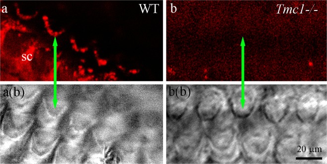
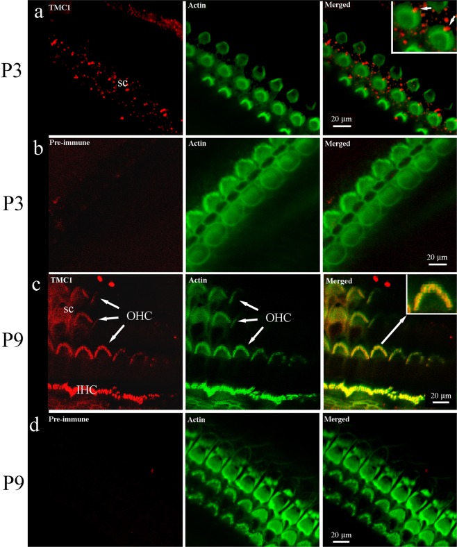
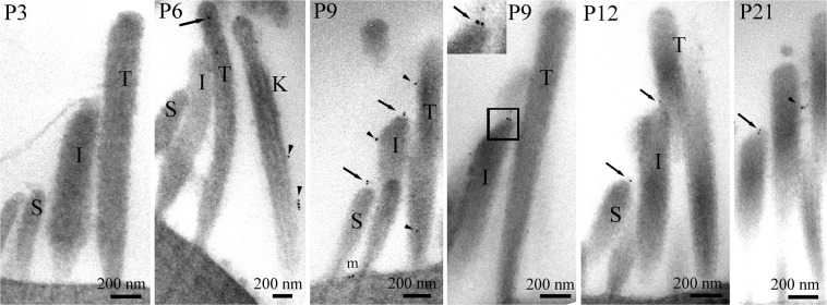
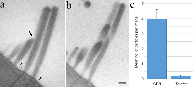
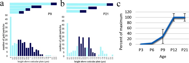
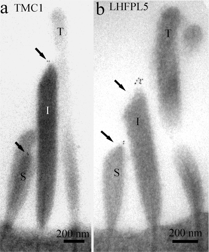
Transmembrane channel like protein 1 (TMC1) is likely to be a pore-forming subunit of the transduction channel of cochlear hair cells that is mechanically gated by tension on tip links in the stereocilia bundle. To localise TMC1 precisely, we labelled mice cochleae of different ages using custom-made polyclonal antibodies to TMC1 for light and transmission electron microscopy (TEM). Immunofluorescence revealed stereocilia labelling at P9 but not at P3 in apical hair cells. Immunogold labelling for TEM confirmed that labelling was absent at P3, and showed weak labelling at P6 with no stereocilia tip labelling, increasing at P9, with specific tip labelling on shorter stereocilia and some throughout the bundle. At P12 and P21, labelling was refined mostly to stereocilia tips. Quantification showed that labelling overall reached maximum by P12, labelling per tip was relatively constant from P9 to P21, but percent tips labelled was reduced from 16% to 8%. Tmc1-/- showed no labelling. Thus TMC1 occurs at the lower end of the tip link, supporting its presence in the MET complex and likely the channel. Tip localisation from P9 onwards coincides with lipoma HMGIC fusion partner-like 5 (LHFPL5), a protein that may be involved in acquiring/maintaining TMC1 localisation.


Conflict of interest statement

The authors declare no competing interests.

Figures

Figure 1
Figure 1
Immunofluorescence labelling and confocal imaging of wild type (WT) and TMC1 knockout (Tmc1−/−) OHC bundles from apical cochlear region at P12. (a) Stereocilia are labelled along with the apices of supporting cells (SC). (b) Neither cell type is labelled in the knockout control. Phalloidin was not used as a counterstain, but bundle localization is evident from the equivalent bright field images a(b) and b(b). Green arrows identify the same bundle in each pair of images.
Figure 2
Figure 2
Immunofluorescent confocal imaging of organ of Corti from the apical cochlear region at P3 and P9. (a) TMC1 is not detected on stereocilia at P3, although the kinocilium appears labelled (arrow in merged image, inset), and speckled labelling is noted on supporting cells. Phalloidin confirms the presence of hair bundles at the same focal plane. (b) Pre-immune serum did not generate any labelling at P3. (c) TMC1 labelling was detected in IHC and OHC bundles at P9; colocalisation with stereocilia is observed as a yellow colour in the merged image. Weak fluorescence was detected on the hair-cell apical surface. The inset shows an enlargement that reveals red and green signal in the indicated (arrow) bundle can also be distinguished. (d) Pre-immune serum did not produce labelling. In all red channel images, the green channel was subtracted using the confocal software to remove bleed through from the phalloidin staining.
Figure 3
Figure 3
Immunogold labelling of OHC stereocilia bundles from apical cochlear regions. Gold particles could not be detected at P3 but were present in small quantities, apparently randomly in the bundle (arrows) and on the kinocilium (arrowheads) at P6. By P9, gold particles were observed on the tips of stereocilia in a position coinciding with the lower end of the tip link (arrows, inset) and occasionally on the shafts (arrowheads). At P12 and P21 tip labelling continued to be present whilst shaft labelling was sparse (arrowheads). Some apical membrane labelling was detected at all ages, as seen for example at P9 (m).
Figure 4
Figure 4
Simultaneous immunogold labelling of (a) wild type and (b) Tmc1−/− OHC bundles from apical cochlear regions, and (c) gold particle counts (mean ± SEM, n = 3) from both at P10. Virtually no labelling was detected on Tmc1−/− bundles whereas labelling on wild type was present as before at shorter stereocilia tips (arrow) and on the apical membrane (arrowheads). The mean gold particles per bundle was significantly different between the two [Wilcoxon (Mann-Whitney) test, p < 0.05].
Figure 5
Figure 5
(a,b) Graphs showing the distribution of particles from the apical membrane to the tips of the tallest stereocilia in OHC bundles from the cochlear apex at P9 and P21. The lengths of each row as determined across ~50 images is shown horizontally above the histogram: dark blue bars represent the tip regions. (a) At P9, particles were found relatively evenly spread through the shorter rows of stereocilia. (b) By P21, labelling was refined to coincide mainly with the tips of short and intermediate stereocilia. (c) Graph showing n/n(max)% particles in approx. 50 images of OHC bundles at different ages. The n/n(max)% is almost 0 at P3 but rises through P9 to reach 100% at P12 which is maintained at P21. The high variance at P9 is indicative of one higher and two lower values.
Figure 6
Figure 6
Comparison of TMC1 (a) and LHFPL5 (b) labelling in OHC bundles from apical cochlear regions at P12. The labelling patterns are similar. Note that in a one short stereocilium is superimposed on a slightly taller one; the particles seen are at the tips of the shorter of these two (arrows).

References

    1. Hackney CM, Furness DN. The composition and role of cross links in mechanoelectrical transduction in vertebrate sensory hair cells. J. Cell Sci. 2013;126:1721–1731. doi: 10.1242/jcs.106120. - DOI - PubMed
    1. Fettiplace R, Hackney CM. The sensory and motor roles of auditory hair cells. Nat. Rev. Neurosci. 2006;7:19–29. doi: 10.1038/nrn1828. - DOI - PubMed
    1. Kazmierczak P, et al. Cadherin 23 and protocadherin 15 interact to form tip-link filaments in sensory hair cells. Nature. 2007;449:87–91. doi: 10.1038/nature06091. - DOI - PubMed
    1. Xiong W, Grillet N, Elledge HM, Wagner HF, Zhao B. TMHS is an integral component of the mechanotransduction machinery of cochlear hair cells. Cell. 2012;151:1283–1295. doi: 10.1016/j.cell.2012.10.041. - DOI - PMC - PubMed
    1. Mahendrasingam S, Fettiplace R, Alagramam KN, Cross E, Furness DN. Spatiotemporal changes in the distribution of LHFPL5 in mice cochlear hair bundles during development and in the absence of PCDH15. PLoS One. 2017;12:e0185285. doi: 10.1371/journal.pone.0185285. - DOI - PMC - PubMed

Publication types