. 2019 Feb 4;9(1):1265.
doi: 10.1038/s41598-018-37666-5.

Oxo-aglaiastatin-Mediated Inhibition of Translation Initiation

Affiliations

Oxo-aglaiastatin-Mediated Inhibition of Translation Initiation

Rayelle Itoua Maïga et al. Sci Rep. .

Abstract

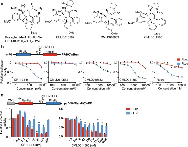
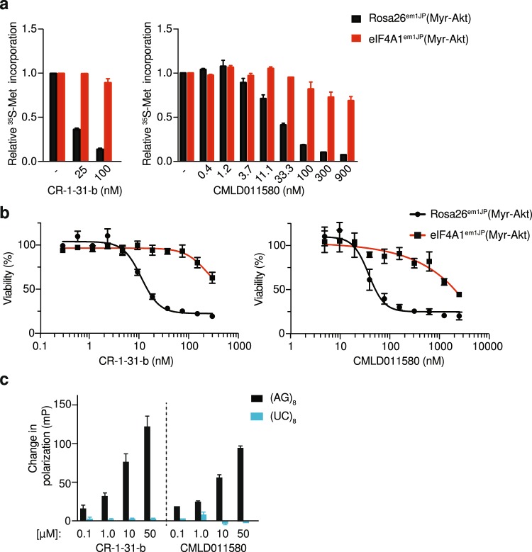
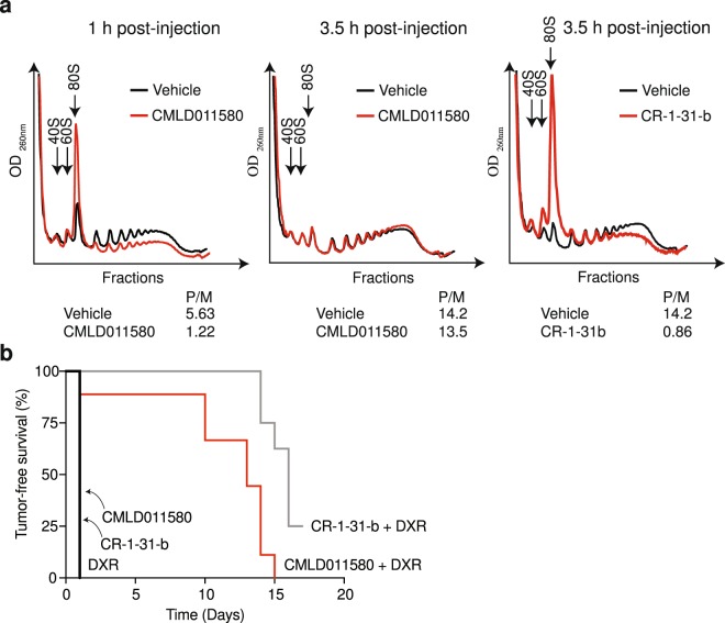
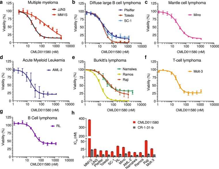
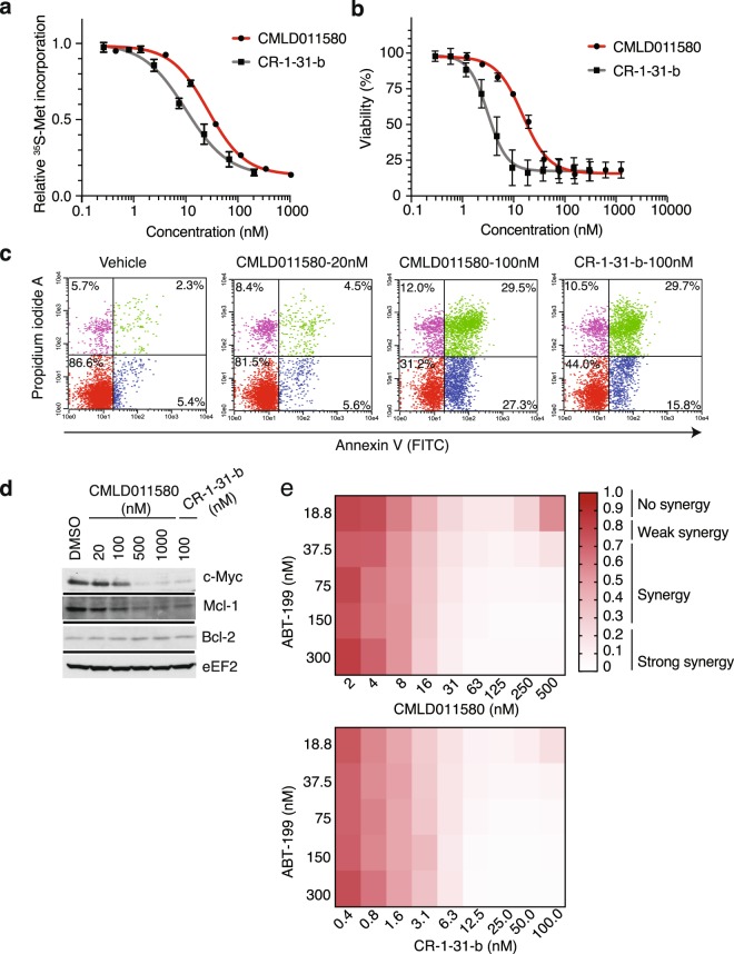
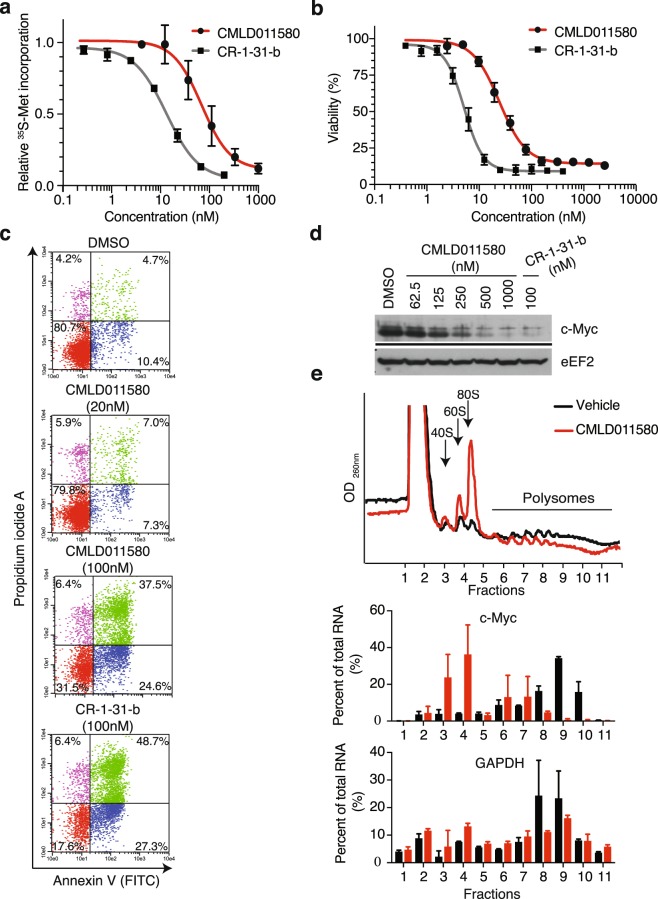
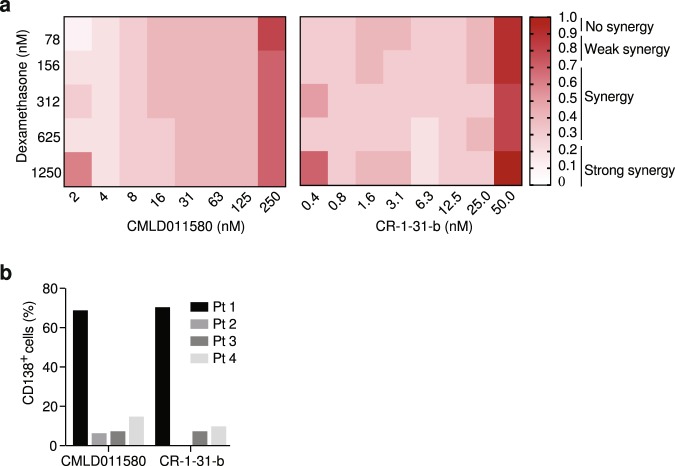
Translation is a highly regulated process that is perturbed in human cancers, often through activation of the PI3K/mTOR pathway which impacts directly on the ribosome recruitment phase of translation initiation. While significant research has focused on "drugging" components of the PI3K/mTOR network, efforts have also been directed towards inhibiting eukaryotic initiation factor (eIF) 4F-dependent translation. Small molecule inhibitors of this complex have been identified, characterized, and used to validate the rationale of targeting this step to curtail tumor cell growth and modulate chemotherapy response. One such class of compounds are the rocaglates, secondary metabolites from the plant genus Aglaia, which target the RNA helicase subunit of eIF4F, eIF4A. Here we explore the ability of synthetic derivatives of aglaiastatins and an aglaroxin derivative to target the translation process in vitro and in vivo and find the synthetic derivative oxo-aglaiastatin to possess such activity. Oxo-aglaiastatin inhibited translation in vitro and in vivo and synergized with doxorubicin, ABT-199 (a Bcl-2 antagonist), and dexamethasone when tested on hematological cancer cells. The biological activity of oxo-aglaiastatin was shown to be a consequence of inhibiting eIF4A1 activity.


Conflict of interest statement

The authors declare no competing interests.

Figures

Figure 1
Figure 1
Aglaiastatin-mediated inhibition of cap-dependent translation. (a) Chemical structure of Roc A, CR-1-31-b, aglaiastatins (CMLD010582, CMLD011580) and aglaroxin (CMLD010833) used in this study. (b) Top: Schematic representation of the FF/HCV/Ren reporter mRNA used herein. Bottom: Assessment of cap- and HCV-mediated translation in the presence of the indicated compound concentrations in Krebs-2 extracts as indicated in the Materials and Methods. Luciferase activity results are expressed relative to values obtained in the presence of DMSO controls. Results are expressed as mean ± SEM of 4 biological replicates. (c) Assessment of CMLD011580 activity in HEK293 cells. Top: Schematic representation of the pcDNA/Ren/HCV/FF expression vector. Bottom: Effect of CMLD011580 on cap-dependent and HCV IRES–mediated translation in HEK293 cells transfected with pcDNA/Ren/HCV/FF. Luciferase activity is expressed relative to values obtained in DMSO-treated cells and is the mean ± SEM of 3 biological replicates.
Figure 2
Figure 2
CMLD011580 targets eIF4A1. (a) Dose-dependent inhibition of protein synthesis in vivo by CMLD011580 is eIF4A1-dependent. Cells were incubated with the indicated concentrations of compound for 1 hour, and [35S]-methionine was added 15 minutes before the end of the incubation. The rate of [35S]-methionine incorporation is expressed relative to that of cells treated with vehicle (DMSO). Results are expressed as the mean ± SEM of 3 biological replicates. (b) Cellular toxicity of CMLD011580 is eIF4A1-dependent. Cells were incubated in the presence of the indicated concentrations of CMLD011580 or (−)-CR-1-31-b for 48 hrs, after which the percentage of viable cells was measured. Results are expressed as the mean ± SEM of 3 biological replicates. (c) Stimulation of eIF4A1:RNA binding by (−)-CR-1-31-b or CMLD011580. Fluorescently labelled RNA (16-mers) containing (AG)8 or (UC)8 repeats were used as probes in these experiments. Shown is the change in fluorescent polarization obtained in the presence of indicated compound concentration, above what was observed in the presence of DMSO. Results are expressed as the mean ± SEM of 3 biological replicates.
Figure 3
Figure 3
In vivo activity of CMLD011580. (a) Assessment of liver polysomes following administration of CMLD011580 in mice. Liver polysomes were isolated following intraperitoneal injection of mice with vehicle or 0.2 mg/kg CMLD011580 or (−)-CR-1-31-b at the indicated time points. (b) Kaplan-Meier plot illustrating the tumor-free survival of Myr-Akt/Eμ-Myc lymphoma bearing mice to the indicated therapeutic regimen. Tumor-free survival was assessed by measuring the time between complete tumor regression following drug administration and the reappearance of tumors in the inguinal lymph nodes. Each cohort had 5–10 mice. p < 0.001 for CMLD011580 + DXR versus DXR.
Figure 4
Figure 4
Dose-dependent inhibition of tumor cell survival in the presence of CMLD011580. (ag) Tumor cells of different hematological origins [multiple myeloma (MM1S, JJN3), diffuse large B cell lymphoma (Pfeiffer, Toledo, SC-1), mantle cell lymphoma (Mino), acute myeloid leukemia (AML-2), Burkitt’s lymphoma (Namalwa, Ramos, Raji), T-cell lymphoma (Molt-3) and B-cell lymphoma (RL)] were incubated with the indicated concentrations of CMLD011580 for 48 hrs and cell viability assessed by CellTiter Glo. Results are expressed relative to vehicle controls and are the mean ± SEM of 3 biological replicates. (h) IC50 of (−)-CR-1-31-b and CMDL011580 towards the indicated cells lines. Results are expressed as mean ± SEM of 3 biological replicates.
Figure 5
Figure 5
Biological properties of CMLD011580 on the THL-DLBCL cell line, SC-1. (a) Dose-dependent inhibition of protein synthesis by (−)-CR-1-31-b and CMLD011580 in SC-1 cells. Cells were incubated with the indicated concentrations of compound for 1 h and [35S]-methionine was added 15 minutes before the end of the incubation. The rate of [35S]-methionine incorporation is expressed relative to that of cells treated with vehicle (DMSO). Results are expressed as mean ± SEM of 3 biological replicates. (b) Relative viability of SC-1 cells exposed to (−)-CR-1-31-b or CMLD011580 for 48 h was assessed by CellTiter Glo. Results are expressed as mean ± SEM of 3 biological replicates. (c) CMLD011580 induces apoptosis in SC-1 cells. Apoptosis was assessed 48 h after compound addition at the indicated concentrations by annexin V and PI staining. Presented is a representative image of 2 independent experiments. (d) Western blot of extracts prepared from SC-1 cells treated with the indicated compounds concentrations for 5 h and probed with antibodies to the indicated proteins. Blots were obtained from the same membrane sequentially probed with the indicated antibodies and are cropped for ease of comparison. (e) Synergistic activity between ABT-199 and CMLD011580 or (−)-CR-1-31-b in SC-1 cells. Cell viability was measured 24 h following incubation with combinations of either of the compounds with ABT-199. n = 3.
Figure 6
Figure 6
Effect of CMLD011580 on multiple myeloma JJN3 cell viability. (a) Dose-dependent inhibition of protein synthesis by (−)-CR-1-31-b and CMLD011580 in JJN3 cells. Cells were incubated with the indicated compound concentrations for 1 h and [35S]-methionine was added 15 minutes before the end of the incubation. The rate of [35S]-methionine incorporation is expressed relative to that of cells treated with vehicle (DMSO). Results are expressed as mean ± SEM of 3 biological replicates. (b) Relative survival of JJN3 cells exposed to (−)-CR-1-31-b or CMLD011580 for 48 h with cell viability assessed by CellTiter Glo. Results are expressed as mean ± SEM of 3 biological replicates. (c) CMLD011580 induces apoptosis in JJN3 cells. Apoptosis was assessed 24 h after compound addition at the indicated concentrations by annexin V and propidium iodide staining. Presented is a representative image of 2 experiments. (d) Western blot of extracts prepared from JJN3 cells treated with the indicated compound concentrations (nM) for 5 h and probed with antibodies to the proteins indicated to the right. Blots were obtained from the same membrane sequentially probed with the indicated antibodies and are cropped for ease of comparison. (e) Polysome analysis of JJN3 cells treated with vehicle (0.05% DMSO) or 25 nM CMLD011580 for 1 h. Polysomes were fractionated on a 10–50% sucrose gradient. The distribution of MYC and GAPDH mRNA within the polysome fractions of vehicle (black boxes) or CMLD011580 (red boxes) treated cells was measured by RT-qPCR. Results are expressed as mean ± SEM of 2 biological replicates.
Figure 7
Figure 7
Biological activity of CMLD011580 in multiple myeloma cells. (a) DEX-dependent synergy with CMLD011580 in JJN3 cells. Cell viability was measured with CellTiter Glo after 48 hours of incubation with different drug combinations. n = 3. (b) Primary human MM cells are sensitive to CMLD011580. Primary tumor cells were exposed ex vivo to 50 nM CMLD011580 or (−)-CR-1-31-b for 72 h, after which point cell viability was determined by flow cytometry as indicated in the Materials and Methods. Presented is the percentage of CD138+ cells surviving drug exposure relative to DMSO controls.

References

    1. Gingras AC, Raught B, Sonenberg N. eIF4 initiation factors: effectors of mRNA recruitment to ribosomes and regulators of translation. Annu Rev Biochem. 1999;68:913–963. doi: 10.1146/annurev.biochem.68.1.913. - DOI - PubMed
    1. Sonenberg N, Hinnebusch AG. Regulation of translation initiation in eukaryotes: mechanisms and biological targets. Cell. 2009;136:731–745. doi: 10.1016/j.cell.2009.01.042. - DOI - PMC - PubMed
    1. Bhat M, et al. Targeting the translation machinery in cancer. Nat Rev Drug Discov. 2015;14:261–278. doi: 10.1038/nrd4505. - DOI - PubMed
    1. Sadlish H, et al. Evidence for a functionally relevant rocaglamide binding site on the eIF4A-RNA complex. ACS Chem Biol. 2013;8:1519–1527. doi: 10.1021/cb400158t. - DOI - PMC - PubMed
    1. Chu J, et al. CRISPR-Mediated Drug-Target Validation Reveals Selective Pharmacological Inhibition of the RNA Helicase, eIF4A. Cell Rep. 2016;15:2340–2347. doi: 10.1016/j.celrep.2016.05.005. - DOI - PMC - PubMed

Publication types

MeSH terms

LinkOut - more resources