. 2019 Feb 4;9(1):1283.
doi: 10.1038/s41598-018-37852-5.

Autoimmune rheumatic disease IgG has differential effects upon neutrophil integrin activation that is modulated by the endothelium

Affiliations

Autoimmune rheumatic disease IgG has differential effects upon neutrophil integrin activation that is modulated by the endothelium

Akif A Khawaja et al. Sci Rep. .

Abstract

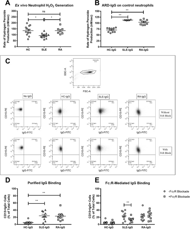
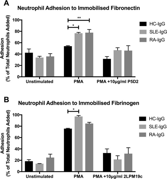
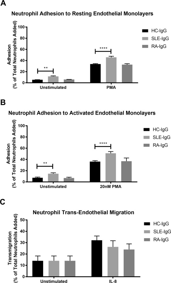
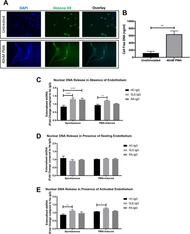
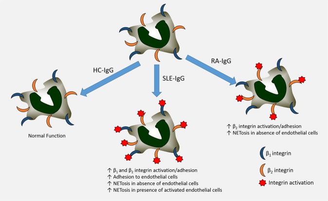
The importance of neutrophils in the pathogenesis of autoimmune rheumatic diseases, such as systemic lupus erythematosus (SLE) and rheumatoid arthritis (RA), is increasingly recognised. Generation of reactive oxygen species (ROS) and release of neutrophil extracellular traps (NETs) by activated neutrophils are both thought to contribute to pathology; although the underlying mechanisms, particularly the effects of IgG autoantibodies upon neutrophil function, are not fully understood. Therefore, we determined whether purified IgG from patients with SLE or RA have differential effects upon neutrophil activation and function. We found that SLE- and RA-IgG both bound human neutrophils but differentially regulated neutrophil function. RA- and SLE-IgG both increased PMA-induced β1 integrin-mediated adhesion to fibronectin, whilst only SLE-IgG enhanced αMβ2 integrin-mediated adhesion to fibrinogen. Interestingly, only SLE-IgG modulated neutrophil adhesion to endothelial cells. Both SLE- and RA-IgG increased ROS generation and DNA externalisation by unstimulated neutrophils. Only SLE-IgG however, drove DNA externalisation following neutrophil activation. Co-culture of neutrophils with resting endothelium prevented IgG-mediated increase of extracellular DNA, but this inhibition was overcome for SLE-IgG when the endothelium was stimulated with TNF-α. This differential pattern of neutrophil activation has implications for understanding SLE and RA pathogenesis and may highlight avenues for development of novel therapeutic strategies.


Conflict of interest statement

The authors declare no competing interests.

Figures

Figure 1
Figure 1
SLE- and RA-IgG both bind neutrophils and prime neutrophils. (A) Neutrophils were isolated from patients with SLE (n = 12) or RA (n = 7) and healthy volunteers (n = 12), and rates of H2O2 production in response to 50 nM PMA using Amplex UltraRed. Data are presented as each point representing a patient sample tested with the mean and SEM also being displayed and were analysed by Kruskal-Wallis test (p = 0.0039) with a Dunn’s multiple comparison test. (B) H2O2 generation in control neutrophils was assessed in the presence of ARD-IgG in the presence of 50 nM PMA. Data are displayed as the mean and SEM from three independent experiments, with each point representing the average of IgG sample over the experimental repeats. Data were analysed by Kruskal-Wallis test (p < 0.0001) with a Dunn’s multiple comparison test. (C) Representative flow cytometry plots examining binding for purified IgG to human neutrophils. Isolated neutrophils were incubated with pooled IgG, composed of 5 different IgG samples in the absence or presence of FcγR blockade, and stained for CD15 and human IgG. IgG binding was determined by quantifying the amount of CD15+IgG+ cell within the neutrophil gate (D) IgG binding experiments were performed using neutrophils from 9 different healthy donors, with mean and SEM being displayed. Statistical significance was tested by a Kruskal-Wallis (p = 0.0010) test with a Dunn’s multiple comparison test. (E) Additional experiments examined pooled IgG binding in the presence of FcγR blockade and the number of CD15+IgG+ cells quantified by flow cytometry. Data are presented as the mean and SEM of nine donors (with each point representing a different neutrophil donor), which were compared to the number of CD15+IgG+ cells previously determined. Data were tested by repeated measures two-way ANOVA a Bonferroni’s multiple comparison test. ns = no significance, *p < 0.05, **p < 0.01.
Figure 2
Figure 2
SLE- IgG enhances both neutrophil β1 and β2 integrin activation. BCECF-AM labelled neutrophil adhesion to immobilised integrin ligands was assessed in the presence of purified IgG to evaluate integrin activation. (A) 20 μg/ml fibronectin was immobilised and neutrophil adhesion examined in the absence or presence of 20 nM PMA and the 10 μg/ml pan-β1 integrin blocking antibody P5D2. (B) 200 μg/ml fibrinogen was immobilised and neutrophil adhesion evaluated in the absence or presence of 20 nM PMA and the 10 μg/ml αMβ2-specifc blocking antibody 2LPM19c. In both cases, data are presented as the mean and SEM from three independent experiments and analysed by two-way ANOVA with a Dunnett’s multiple comparison test. *p < 0.05, **p < 0.01.
Figure 3
Figure 3
SLE- IgG increased neutrophil adhesion to endothelial cells, but not trans-endothelial migration. BCECF-AM labelled neutrophil adhesion was assessed in the absence or presence of purified IgG and 20 nM PMA to (A) resting endothelial cells and (B) endothelial cells that had been activated with TNF-α prior to experimentation. (C) Fluorescently labelled neutrophil trans-endothelial migration was determined in the absence or presence of purified IgG and 150 ng/ml IL-8, using a trans-well cell culture system and flow cytometry. Cells were quantified by comparing enumerated cells in the lower chamber to the total cells added. In both cases, data are displayed as the mean and SEM of three independent experiments and were tested for significance using two-way ANOVA with a Dunnett’s multiple comparison test. **p < 0.01, ****p < 0.0001.
Figure 4
Figure 4
SLE- IgG increased neutrophil NETosis in the presence of endothelial cells. (A) PMA-induced NETosis was confirmed by immunofluorescence. Neutrophils were incubated on coverslips in the absence or presence of 40 nM PMA and then fixed. Coverslips were then stained for histone H3 and mounted using a DAPI-mounting medium. (B) Cell supernatants from neutrophils cultured in the absence or presence of 40 nM were analysed using the Quanti-iT PicoGreen dsDNA kit. Data are presented as the mean and SEM of three independent experiments. Statistical significance was determined by a Mann-Whitney test. (CE) IgG-mediated extracellular DNA release was assessed in (C) the absence of an endothelial monolayer; (D) the presence of resting endothelial cells; and (E) the presence of activated endothelial cells. Neutrophils were cultured for 4 hours in the absence or presence of 40 nM PMA, after which cell supernatants were assessed for cell-free dsDNA. For endothelial cell activation, HUVEC were pre-stimulated with 10 ng/ml TNF-α for 24 hours and washed with warmed PBS prior to addition of neutrophils. Data are presented as the mean and SEM of three independent experiments and analysed using a two-way ANOVA with a Dunnet’s multiple comparison test. *p < 0.05, **p < 0.01, ****p < 0.0001.
Figure 5
Figure 5
Schematic of overall findings. SLE- and RA-IgG displayed differential effects upon HC-neutrophils in our experiments. SLE-IgG enhanced both β1 and β2 integrin-mediated adhesion and also adhesion to endothelial cells. In contrast, RA-IgG only increased β2 integrin-mediated adhesion. We observed higher levels of NETosis both in absence and presence of activated endothelial cells with the addition of SLE-IgG, whilst RA-IgG only enhanced NETosis in the absence of an endothelial monolayer.

Similar articles

Cited by

References

    1. Nemeth T, Mocsai A. The role of neutrophils in autoimmune diseases. Immunology letters. 2012;143:9–19. doi: 10.1016/j.imlet.2012.01.013. - DOI - PubMed
    1. Jorch SK, Kubes P. An emerging role for neutrophil extracellular traps in noninfectious disease. Nature medicine. 2017;23:279–287. doi: 10.1038/nm.4294. - DOI - PubMed
    1. Brinkmann V, et al. Neutrophil extracellular traps kill bacteria. Science. 2004;303:1532–1535. doi: 10.1126/science.1092385. - DOI - PubMed
    1. Gupta AK, et al. Activated endothelial cells induce neutrophil extracellular traps and are susceptible to NETosis-mediated cell death. FEBS letters. 2010;584:3193–3197. doi: 10.1016/j.febslet.2010.06.006. - DOI - PubMed
    1. Carmona-Rivera C, Zhao W, Yalavarthi S, Kaplan MJ. Neutrophil extracellular traps induce endothelial dysfunction in systemic lupus erythematosus through the activation of matrix metalloproteinase-2. Annals of the rheumatic diseases. 2015;74:1417–1424. doi: 10.1136/annrheumdis-2013-204837. - DOI - PMC - PubMed

Publication types

MeSH terms