

Decreased dopamine in striatum and difficult locomotor recovery from MPTP insult after exposure to radiofrequency electromagnetic fields
- PMID: 30718744
- PMCID: PMC6362053
- DOI: 10.1038/s41598-018-37874-z
Decreased dopamine in striatum and difficult locomotor recovery from MPTP insult after exposure to radiofrequency electromagnetic fields
Abstract
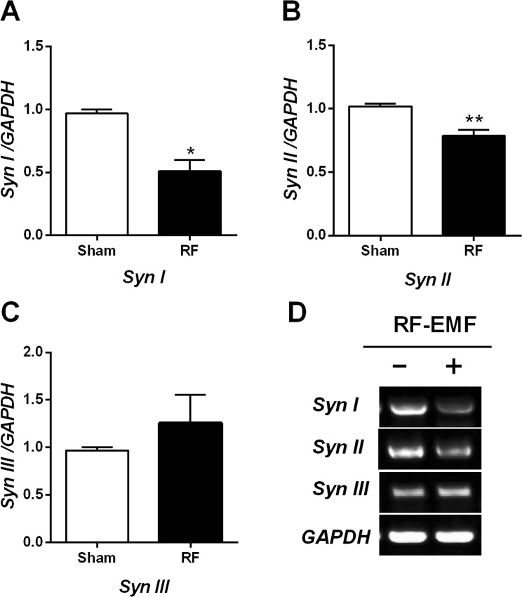
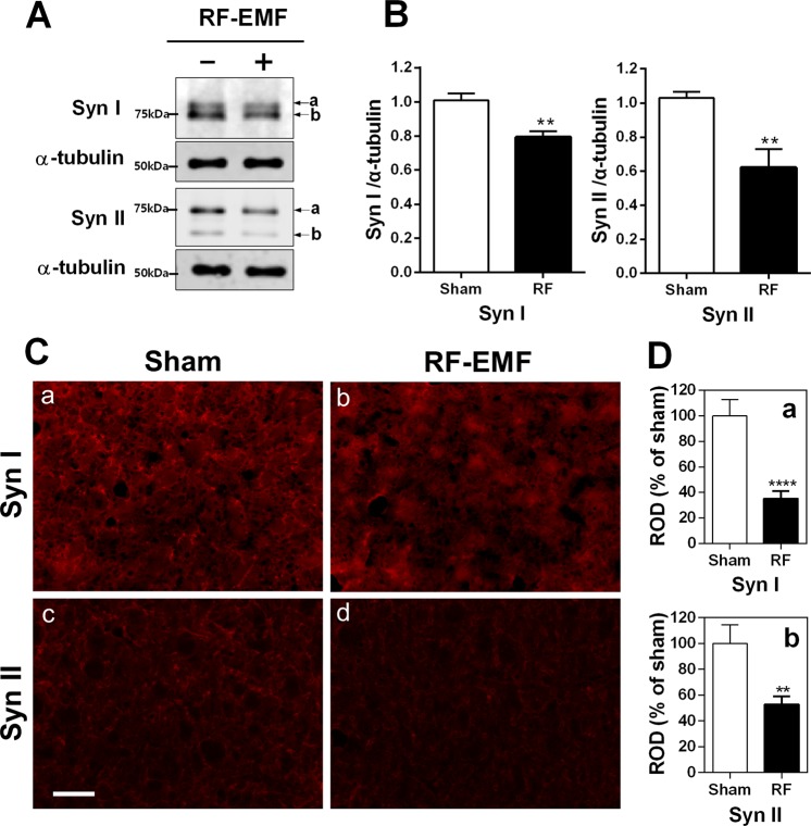
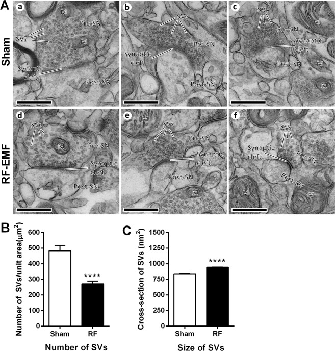
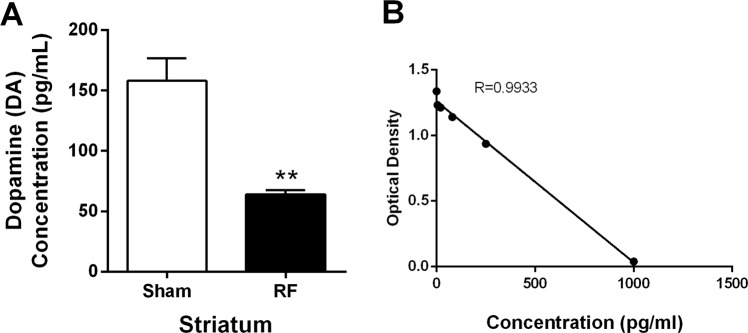
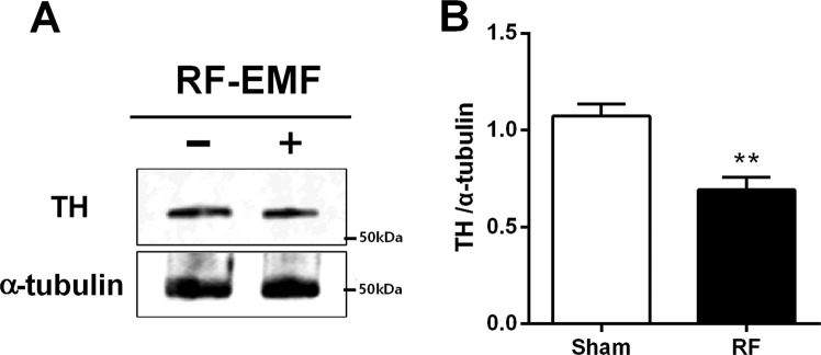
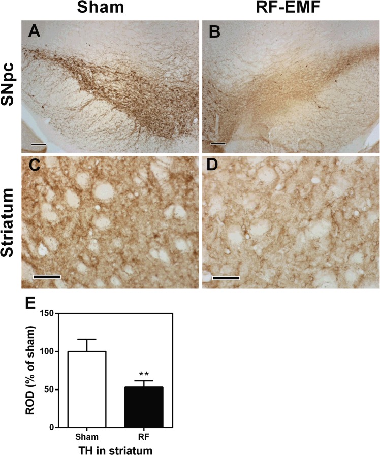
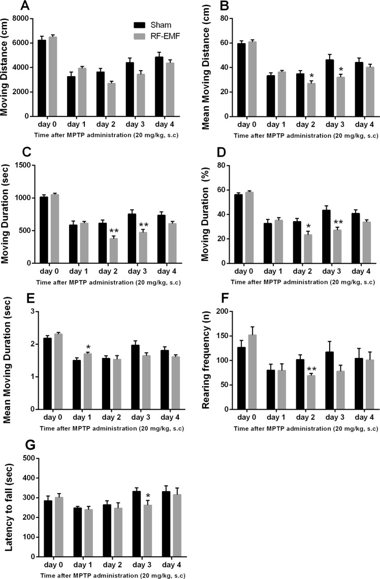
Concern is growing about possible neuronal effects of human exposure to radiofrequency electromagnetic fields because of the increasing usage of cell phones and the close proximity of these devices to the brain when in use. We found that exposure to a radiofrequency electromagnetic field (RF-EMF) of 835 MHz (4.0 W/kg specific absorption rate [SAR] for 5 h/day for 12 weeks) affects striatal neurons in C57BL/6 mice. The number of synaptic vesicles (SVs) in striatal presynaptic boutons was significantly decreased after RF-EMF exposure. The expression levels of synapsin I and II were also significantly decreased in the striatum of the RF-EMF-exposed group. RF-EMF exposure led to a reduction in dopamine concentration in the striatum and also to a decrease in the expression of tyrosine hydroxylase in striatal neurons. Furthermore, in behavioral tests, exposure to RF-EMF impeded the recovery of locomotor activities after repeated treatments with 1-methyl-4-phenyl-1,2,3,6-tetrahydropyridine (MPTP). These results suggest that the observed decrease in dopamine concentration in the striatum was caused by both a reduction in the number of dopaminergic neurons and a decline in the number of SVs. The decreased dopamine neuron numbers and concentration seen after RF-EMF exposure would have caused the difficult recovery after MPTP treatment. In summary, our results strongly suggest that exposing the brain to RF-EMF can decrease the number of SVs and dopaminergic neurons in the striatum. These primary changes impair the recovery of locomotor activities following MPTP damage to the striatum.
Conflict of interest statement
The authors declare no competing interests.
Figures
Similar articles
-
Exposure to RF-EMF Alters Postsynaptic Structure and Hinders Neurite Outgrowth in Developing Hippocampal Neurons of Early Postnatal Mice.Int J Mol Sci. 2021 May 19;22(10):5340. doi: 10.3390/ijms22105340. Int J Mol Sci. 2021. PMID: 34069478 Free PMC article.
-
Changes in numbers and size of synaptic vesicles of cortical neurons induced by exposure to 835 MHz radiofrequency-electromagnetic field.PLoS One. 2017 Oct 18;12(10):e0186416. doi: 10.1371/journal.pone.0186416. eCollection 2017. PLoS One. 2017. PMID: 29045446 Free PMC article.
-
Trafficking of synaptic vesicles is changed at the hypothalamus by exposure to an 835 MHz radiofrequency electromagnetic field.Gen Physiol Biophys. 2019 Sep;38(5):379-388. doi: 10.4149/gpb_2019020. Epub 2019 Aug 14. Gen Physiol Biophys. 2019. PMID: 31411574
-
Effects of radiofrequency electromagnetic field (RF-EMF) exposure on male fertility: A systematic review of experimental studies on non-human mammals and human sperm in vitro.Environ Int. 2024 Mar;185:108509. doi: 10.1016/j.envint.2024.108509. Epub 2024 Feb 19. Environ Int. 2024. PMID: 38492496
-
Threshold of radiofrequency electromagnetic field effect on human brain.Int J Radiat Biol. 2021;97(11):1505-1515. doi: 10.1080/09553002.2021.1969055. Epub 2021 Aug 23. Int J Radiat Biol. 2021. PMID: 34402382 Review.
Cited by
-
Comparison of pulsed and continuous electromagnetic field generated by WPT system on human dermal and neural cells.Sci Rep. 2024 Mar 6;14(1):5514. doi: 10.1038/s41598-024-56051-z. Sci Rep. 2024. PMID: 38448548 Free PMC article.
-
Exposure to RF-EMF Alters Postsynaptic Structure and Hinders Neurite Outgrowth in Developing Hippocampal Neurons of Early Postnatal Mice.Int J Mol Sci. 2021 May 19;22(10):5340. doi: 10.3390/ijms22105340. Int J Mol Sci. 2021. PMID: 34069478 Free PMC article.
-
1800 MHz Radiofrequency Electromagnetic Field Impairs Neurite Outgrowth Through Inhibiting EPHA5 Signaling.Front Cell Dev Biol. 2021 Apr 12;9:657623. doi: 10.3389/fcell.2021.657623. eCollection 2021. Front Cell Dev Biol. 2021. PMID: 33912567 Free PMC article.
-
Localisation of clozapine during experimental autoimmune encephalomyelitis and its impact on dopamine and its receptors.Sci Rep. 2021 Feb 3;11(1):2966. doi: 10.1038/s41598-021-82667-6. Sci Rep. 2021. PMID: 33536582 Free PMC article.
-
Metformin Protects From Rotenone-Induced Nigrostriatal Neuronal Death in Adult Mice by Activating AMPK-FOXO3 Signaling and Mitigation of Angiogenesis.Front Mol Neurosci. 2020 Jun 18;13:84. doi: 10.3389/fnmol.2020.00084. eCollection 2020. Front Mol Neurosci. 2020. PMID: 32625061 Free PMC article.
References
-
- Wyde, M. et al. Report of Partial findings from the National Toxicology Program Carcinogenesis Studies of Cell Phone Radiofrequency Radiation in Hsd: Sprague Dawley® SD rats (Whole Body Exposure). bioRxiv, 10.1101/055699 (2018).
Publication types
MeSH terms
Substances
LinkOut - more resources
Full Text Sources
Medical
Miscellaneous
