. 2019 May;26(5):151-164.
doi: 10.1038/s41434-019-0058-7. Epub 2019 Feb 4.

Measles vector as a multigene delivery platform facilitating iPSC reprogramming

Affiliations

Measles vector as a multigene delivery platform facilitating iPSC reprogramming

Qi Wang et al. Gene Ther. 2019 May.

Abstract

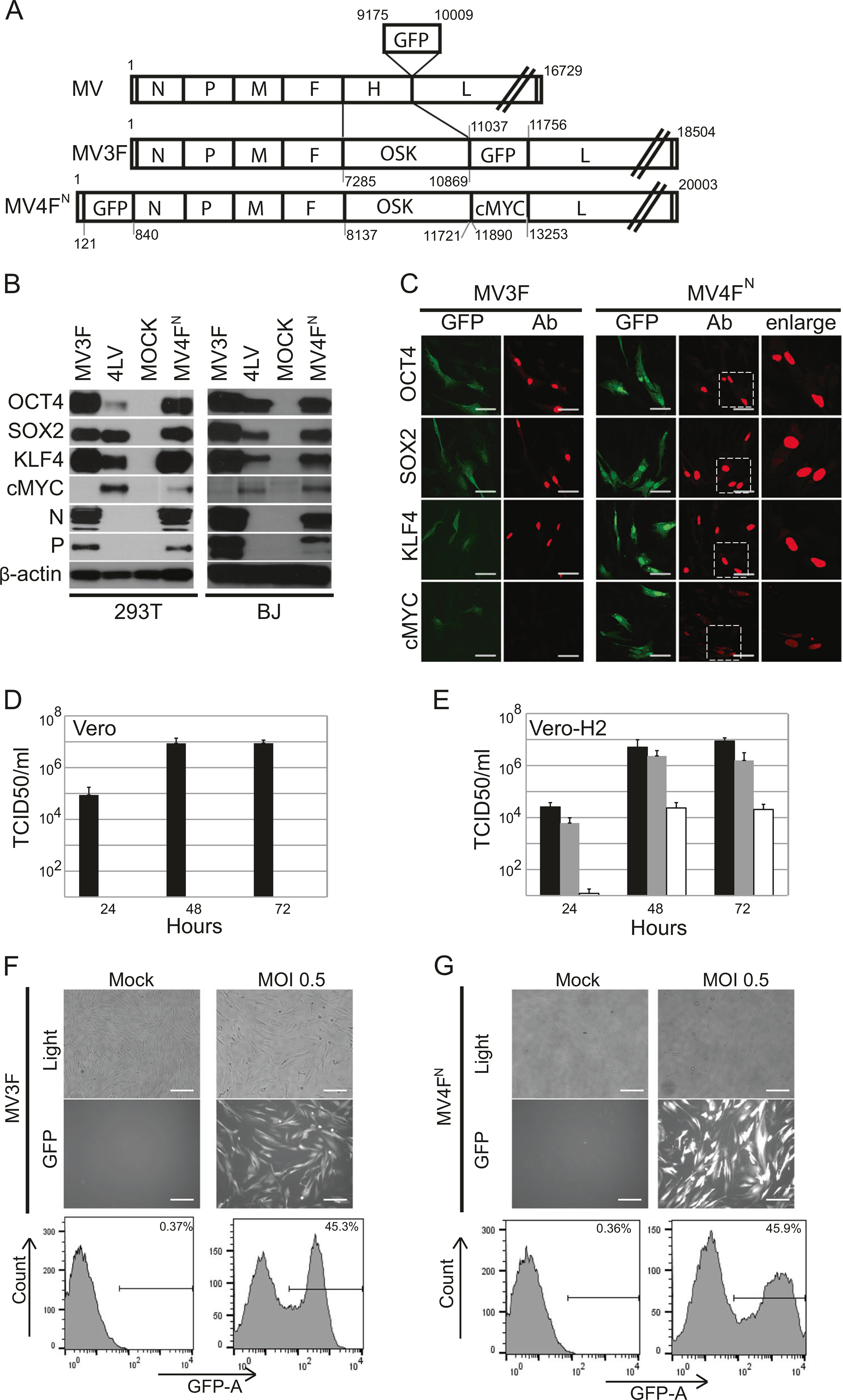
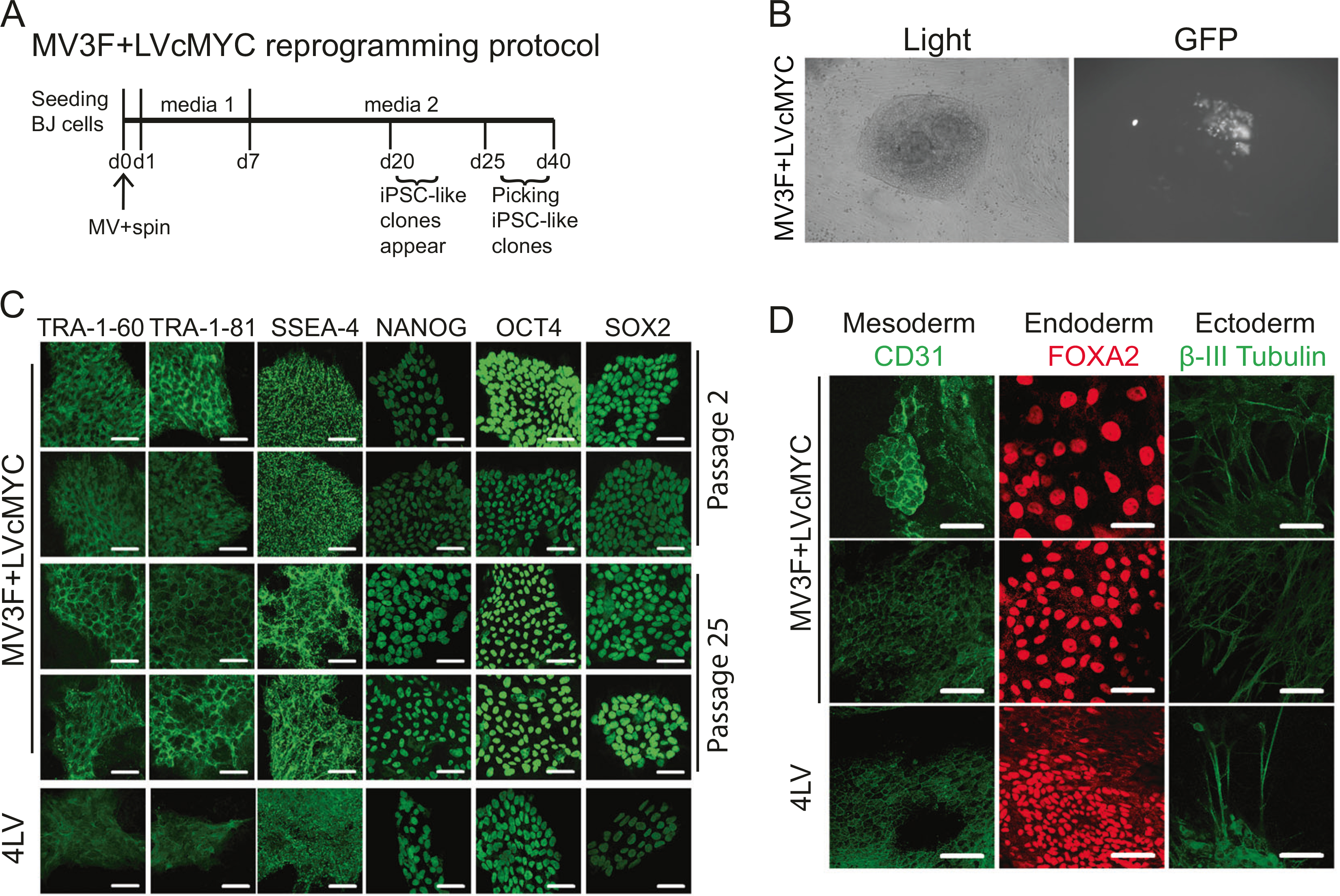
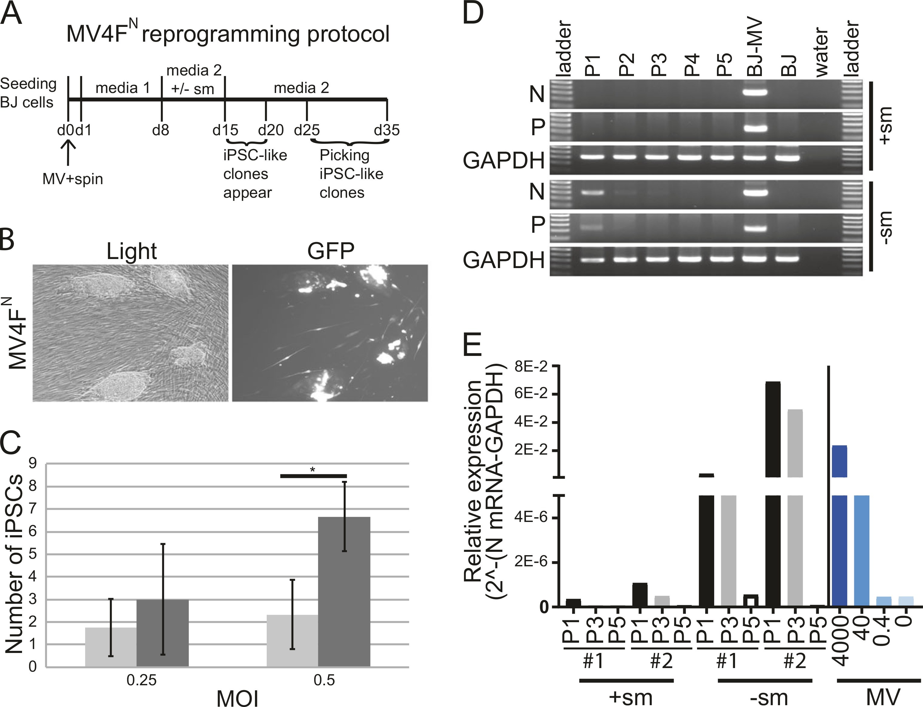
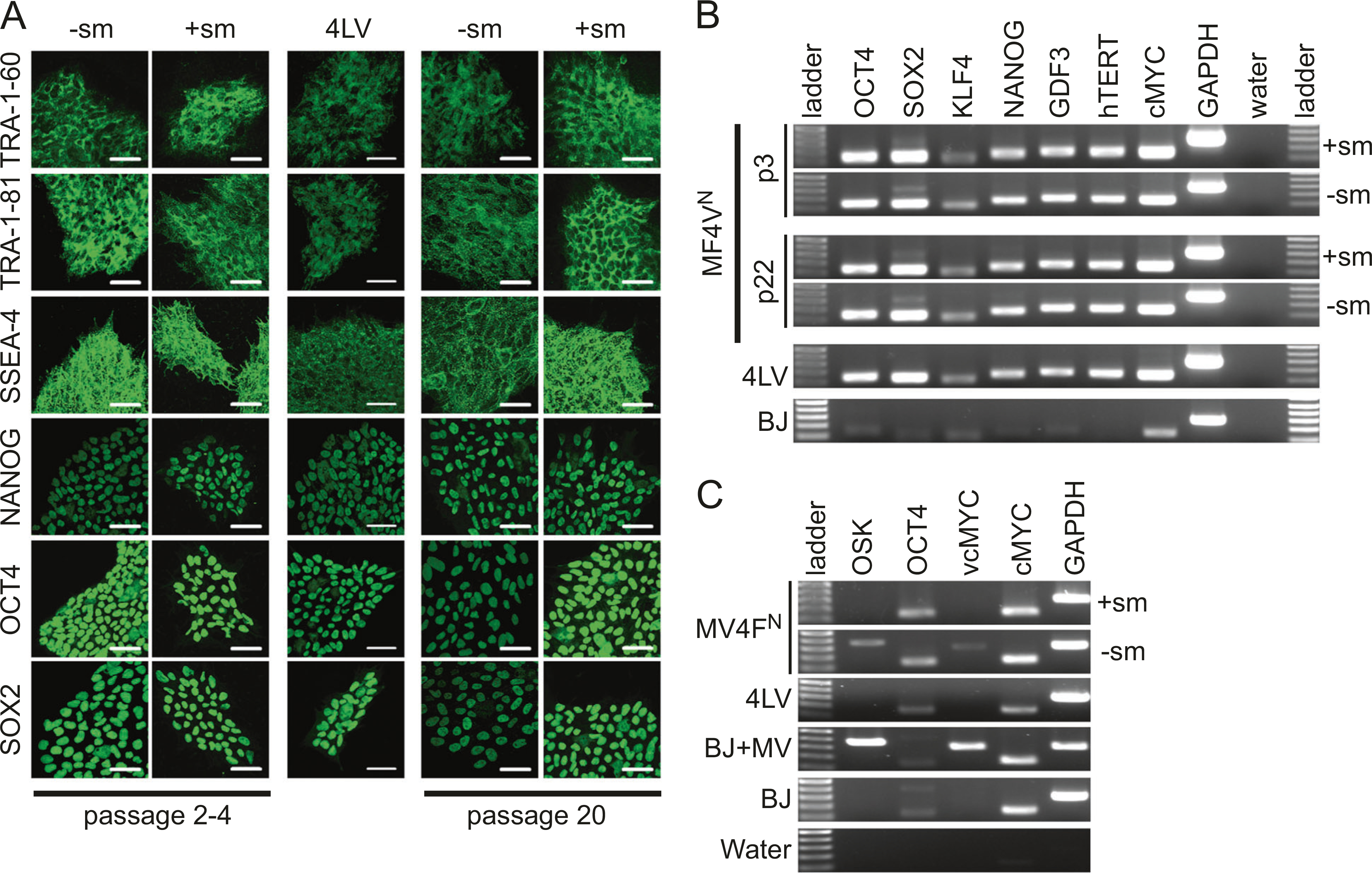
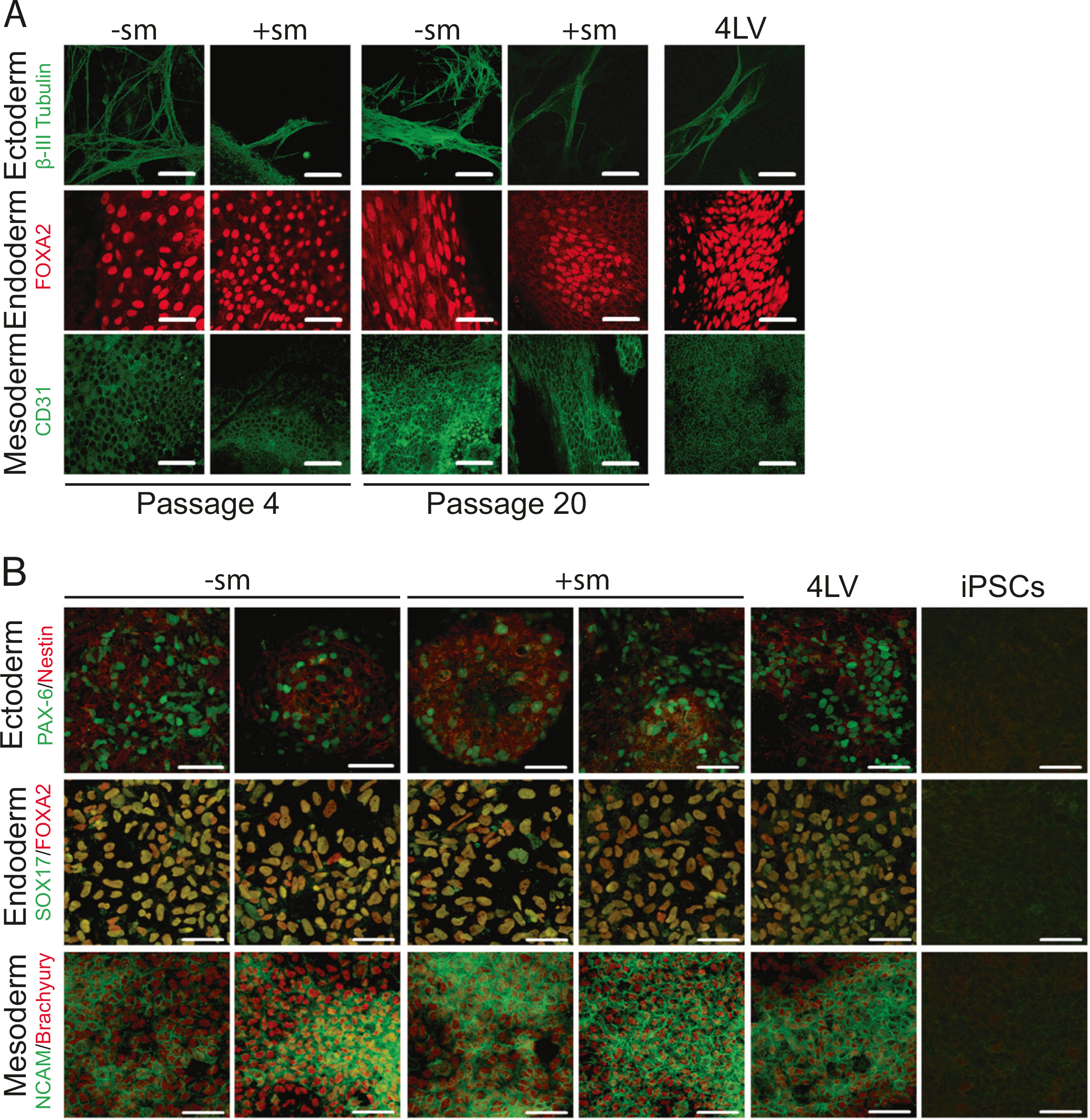
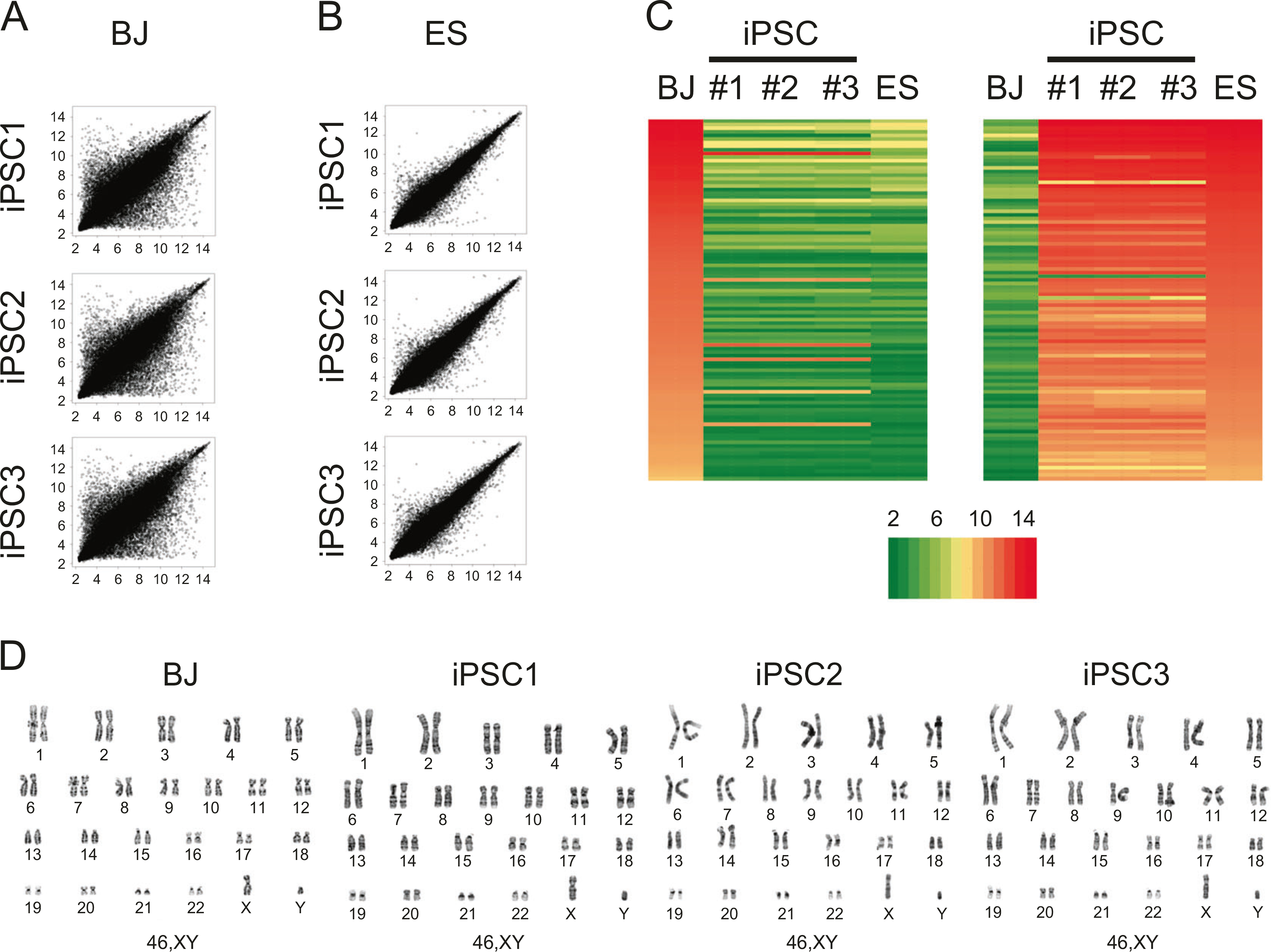
Induced pluripotent stem cells (iPSCs) provide a unique platform for individualized cell therapy approaches. Currently, episomal DNA, mRNA, and Sendai virus-based RNA reprogramming systems are widely used to generate iPSCs. However, they all rely on the use of multiple (three to six) components (vectors/plasmids/mRNAs) leading to the production of partially reprogrammed cells, reducing the efficiency of the systems. We produced a one-cycle measles virus (MV) vector by substituting the viral attachment protein gene with the green fluorescent protein (GFP) gene. Here, we present a highly efficient multi-transgene delivery system based on a vaccine strain of MV, a non-integrating RNA virus that has a long-standing safety record in humans. Introduction of the four reprogramming factors OCT4, SOX2, KLF4, and cMYC via a single, "one-cycle" MV vector efficiently reprogrammed human somatic cells into iPSCs, whereas MV vector genomes are rapidly eliminated in derived iPSCs. Our MV vector system offers a new reprogramming platform for genomic modification-free iPSCs amenable for clinical translation.


Conflict of interest statement

Conflict of interest PD and YI are inventors on a patent application (WO2018064460A1) for the content of the manuscript. The remaining authors declare that they have no conflict of interest.

Figures

Fig. 1
Fig. 1
Generation and characterization of a one-cycle measles virus vector expressing OCT4, SOX2, KLF4, cMYC, and GFP. a Schematic representation of MV(GFP)H (top), MV3F (middle), and MV4FΝ (bottom). The measles virus (MV) antigenome (plus strand) is represented with its 5′ end on the left; the six genes are indicated by capital letters, numbers represent nucleotides position with number 1 being the first nucleotide of measles genome. b Immunoblot analysis of OCT4, SOX2, KLF4, cMYC expression in 293T and human fibroblast (BJ) transduced cells with the indicated vector. Antibodies against the indicated proteins were used. Uninfected BJ and 293T cells (MOCK) were used as controls. Cells transduced with LVOCT4, LVSOX2, LVKLF4, and LVcMYC (4LV) were used as positive control. β-Actin was used as loading control. c Immunofluorescence analysis of OCT4, SOX2, KLF4, cMYC expression in transduced human fibroblast (BJ) cells with the indicated vector. Cells were stained with indicated antibodies (red). GFP (green) was expressed during infection. Scale bars represent 50 μm. d, e Titers of cell-associated and released virus produced upon infection of Vero (d) and Vero-H2 (e) cells with MV3F (gray columns), MV4FN (white columns) or MV (black columns), determined at 24 h, 48 h or 72 h post-infection. Values and error bars reflect the mean and standard deviation of at least two biological replicates. f Level of transduction of human fibroblasts with MV3F. Cells (BJ) were infected with MV3F or mock infected. Forty-eight hours post-infection, pictures were taken under phase contrast (top panels), fluorescence (bottom panels) or quantified by flow cytometry (bottom panel). g Level of transduction of human fibroblasts with MV4FN. Cells (BJ) were infected with MV4FN or mock infected. Forty-eight hours post-infection, pictures were taken under phase contrast (top panels), fluorescence (middle panels) or quantified by flow cytometry (bottom panel). Scale bars represent 0.2 mm
Fig. 2
Fig. 2
Generation of induced pluripotent stem cell (iPSC) using MV3F and elimination of the vector after reprogramming (a). Reprogramming schedule of human fibroblasts (BJ) transduced with MV3F + LVcMYC (MV3F). b Representative iPSC-like clone obtained 20 days post-transduction with MV3F + LVcMYC under light and fluorescence microscopy (left and right panel, respectively). c Two representative MV3F + LVcMYC-derived iPSC clones and one 4LV-derived iPSC control (4LV) were cultured under feeder-free conditions on a matrigel-based slide and examined for expression of human pluripotent stem cell markers by immunofluorescence. Passages 2 and 25 were analyzed. Scale bars represent 50 μm. d The same iPSC clones were analyzed by immunofluorescence for lineage markers for three germ layers (endoderm, mesoderm, and ectoderm). iPSC clones were spontaneously differentiated through embryoid body formation. Pluripotency of derived iPSC clones was verified by generation of cells of ectoderm (β-III tubulin, green, top row), endoderm (FOXA2, red, second row), and mesoderm (CD31, green, bottom row) upon spontaneous differentiation. Clones were tested at passages 4 and 20. Scale bars indicate 50 μm
Fig. 3
Fig. 3
Generation of induced pluripotent stem cell (iPSC) using MV4FN and elimination of the vector after reprogramming (a). Reprogramming schedule of human fibroblasts (BJ) transduced with MV4FN. b Representative pictures of iPSC-like clones obtained ~15 days post-transduction with MV4FN under light and fluorescence microscopy. c Reprogramming efficiency: average number of iPSC clones produced after transduction of 2.1 × 10⁵ BJ cells with MV4FN, with (black columns) or without (gray columns) small molecules. Values and error bars reflect the mean and standard deviation of three biological replicates. *P < 0.05 with small molecules versus without small molecules, Student’s t-test. d Loss of viral gene expression after passaging. Nucleoprotein (N) and Phosphoprotein (P) mRNA expression levels were analyzed in MV4FN -derived iPSC clones by semiquantitative RT-PCR at passages 1, 2, 3, 4, and 5. GAPDH is the cellular internal control, and water is the negative control. Controls: (BJ-MV) BJ cells infected with MV4FN, (BJ) BJ mock infected. e Elimination of the vector analysis. Quantitative RT-PCR analysis of the relative expression of the N mRNA in four iPSC clones obtained in presence ( + sm) or absence (−sm) of small molecules at passage 1, 3, and 5 (P1, P3, and P5; black, gray, and white columns, respectively). The right part of the graph (blue histograms) represents a quantitative PCR from 4000 to 0 molecules vector cDNA genome
Fig. 4
Fig. 4
Characterization of MV4FN-derived induced pluripotent stem cell (iPSC) clones for pluripotency markers. a One representative MV4FN-derived iPSC clones obtained with or without small molecules ( + sm, −sm) and one 4LV-derived iPSC control (4LV) were cultured under feeder-free conditions on a matrigel-based slide and examined for expression of human pluripotent stem cell markers by immunofluorescence. Passages 2–4 and 20 were analyzed. Scale bars represent 50 μm. b RT-PCR analysis assessing transcription of key pluripotency-associated genes (OCT4, SOX2, KLF4, NANOG, GDF3, hTERT, cMYC) using total cellular RNA of two representative iPSC clones obtained with or without small molecules at passages 3 and 22. GAPDH is the cellular internal control, and water is the negative control. c RT-PCR analysis assessing transcription of viral and cellular genes OCT4, and cMYC were analyzed using total cellular RNA of one representative iPSC clones obtained with or without small molecules at passages 1. GAPDH is the cellular internal control, and water is the negative control. BJ cells and measles virus (MV)-infected BJ cells were used as negative and positive control of viral OCT4 and cMYC
Fig. 5
Fig. 5
Spontaneous and guided differentiation of the MV4FN-derived induced pluripotent stem cell (iPSC) clones. a Representative MV4FN-derived iPSC clones obtained with or without small molecules (+sm, −sm) and one 4LV-derived iPSC control (4LV) were analyzed by immunofluorescence for lineage markers for three germ layers (endoderm, mesoderm, and ectoderm). iPSC clones were spontaneously differentiated through embryoid body formation. Pluripotency of derived iPSC clones was verified by generation of cells of ectoderm (β-III tubulin, green, top row), endoderm (FOXA2, red, second row), and mesoderm (CD31, green, bottom row) upon spontaneous differentiation. Clones were tested at passages 4 and 20. Scale bars indicate 50 μm. b Representative MV4FN-derived iPSC clones obtained with or without small molecules (+sm, −sm), and one 4LV-derived iPSC control (4LV) were analyzed by immunofluorescence for lineage markers for three germ layers (endoderm, mesoderm, and ectoderm). iPSC clones were differentiated through guided differentiation using the STEMdiff™ Trilineage Differentiation kit. Pluripotency of derived iPSC clones was verified by generation of cells of ectoderm (Nestin [red] and PAX-6 [green], top row), endoderm (FOXA2 [red] and SOX17 [green], second row), and mesoderm (NCAM [green] and Brachyury [red], bottom row) upon guided differentiation. Clones were tested at passage 4. Control staining was done on not differentiated iPSCs (right three panels, iPSCs). Scale bars indicate 50 μm
Fig. 6
Fig. 6
Global gene expression comparison between measles virus (MV)-derived induced pluripotent stem cell (iPSC) and human ES cells. a Global gene expression patterns were compared between human fibroblast (BJ) and three MV4FN-iPSC clones. b Global gene expression patterns were compared between human embryonic stem (ES) cells (H9, GSM551202) and the same three MV4FN-iPSC clones (GSE122790). c Heat map analysis of human fibroblast (BJ), MV4FN-iPSC clones (iPSC1, iPSC2, and iPSC3) and human ES cells (ES). Expression of genes that are differentially expressed between BJ and iPSC clones. (Right) Top hundred human ES cell-enriched probe sets; (left) top hundred fibroblast enriched probe sets. The color key indicates the color code gene expression in log2 scale. d G-banding chromosome analysis of parental BJ cells and the three same MV4FN-iPSC clones, iPSC1, iPSC2, and iPSC3

References

    1. Takahashi K, Tanabe K, Ohnuki M, Narita M, Ichisaka T, Tomoda K, et al. Induction of pluripotent stem cells from adult human fibroblasts by defined factors. Cell. 2007;131:861–72. - PubMed
    1. Takahashi K, Yamanaka S. Induction of pluripotent stem cells from mouse embryonic and adult fibroblast cultures by defined factors. Cell. 2006;126:663–76. - PubMed
    1. Yu J, Vodyanik MA, Smuga-Otto K, Antosiewicz-Bourget J, Frane JL, Tian S, et al. Induced pluripotent stem cell lines derived from human somatic cells. Science. 2007;318:1917–20. - PubMed
    1. Shi Y, Inoue H, Wu JC, Yamanaka S. Induced pluripotent stem cell technology: a decade of progress. Nat Rev Drug Discov. 2017;16:115–30. - PMC - PubMed
    1. Hu C, Li L. Current reprogramming systems in regenerative medicine: from somatic cells to induced pluripotent stem cells. Regen Med. 2016;11:105–32. - PubMed

Publication types