. 2019 Feb 4;9(1):1144.
doi: 10.1038/s41598-018-38171-5.

Epithelial to mesenchymal transition is mediated by both TGF-β canonical and non-canonical signaling during axolotl limb regeneration

Affiliations

Epithelial to mesenchymal transition is mediated by both TGF-β canonical and non-canonical signaling during axolotl limb regeneration

Fadi Sader et al. Sci Rep. .

Erratum in

Abstract

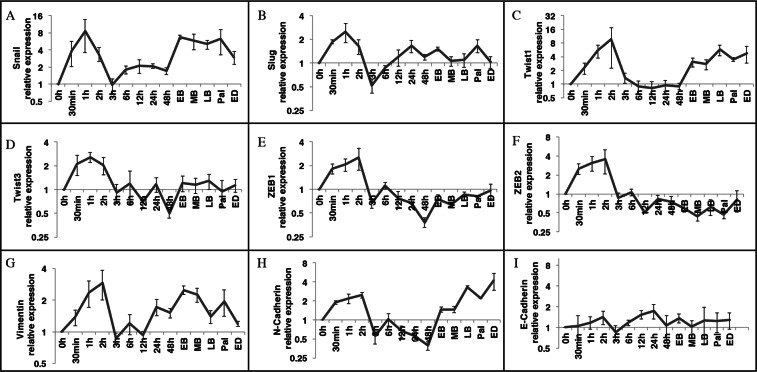
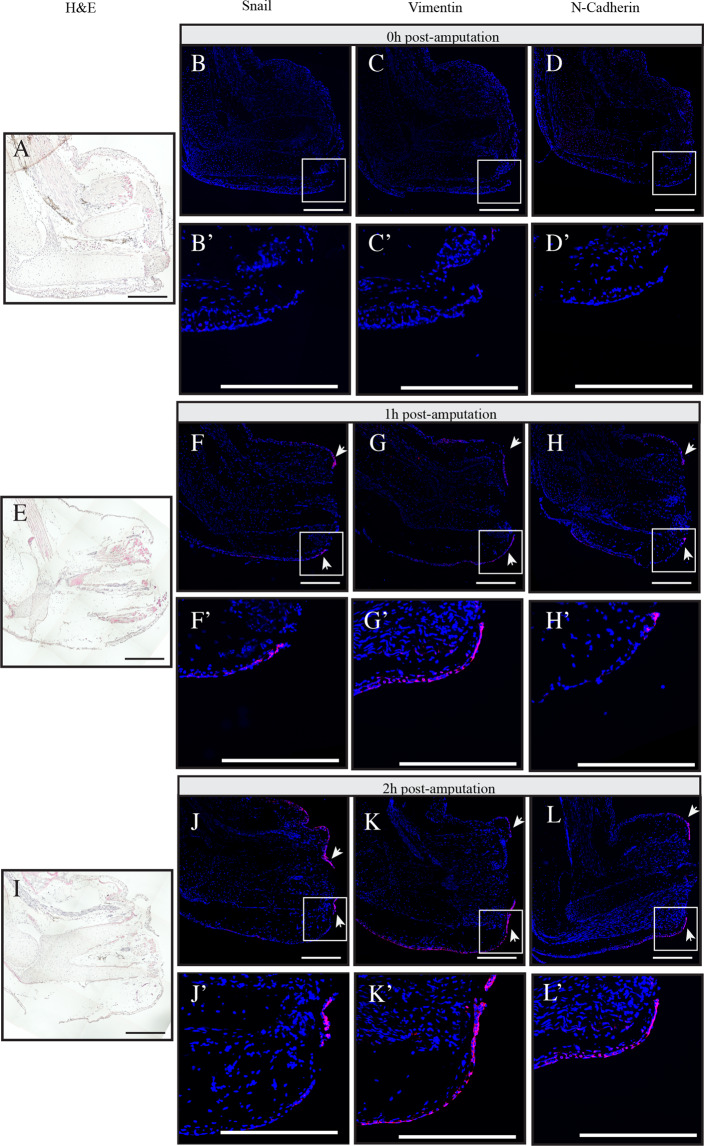
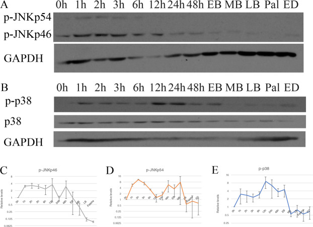
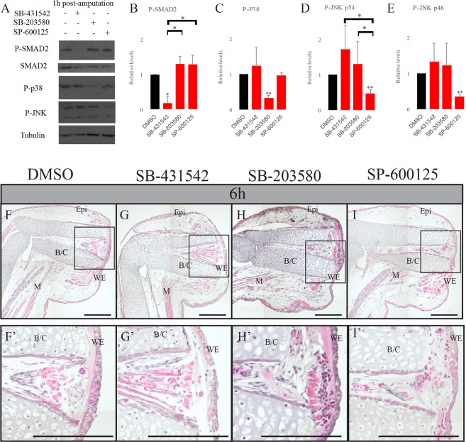
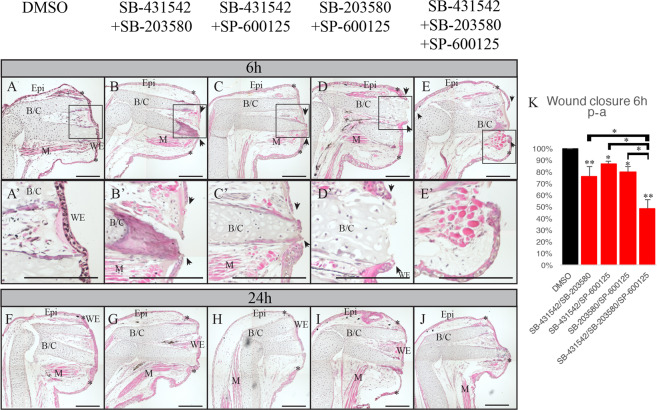
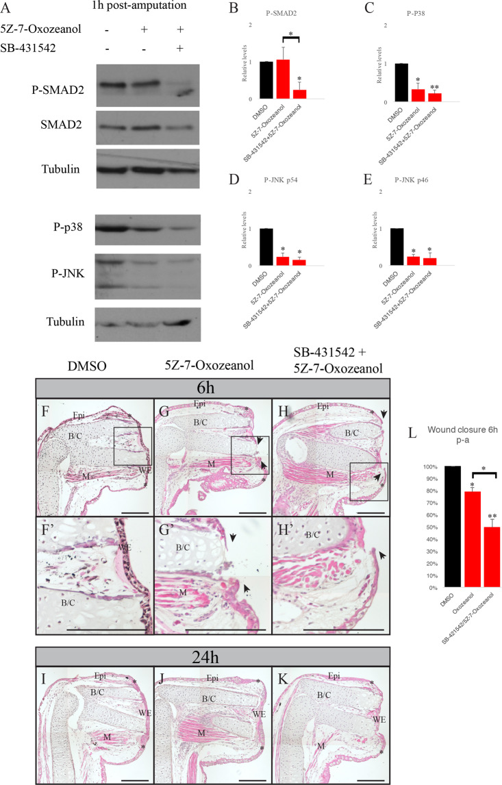
Axolotls have the amazing ability to regenerate. When compared to humans, axolotls display a very fast wound closure, no scarring and are capable to replace lost appendages perfectly. Understanding the signaling mechanism leading to this perfect healing is a key step to help develop regenerative treatments for humans. In this paper, we studied cellular pathways leading to axolotl limb regeneration. We focus on the wound closure phase where keratinocytes migrate to close the lesion site and how epithelial to mesenchymal transitions are involved in this process. We observe a correlation between wound closure and EMT marker expression. Functional analyses using pharmacological inhibitors showed that the TGF-β/SMAD (canonical) and the TGF-β/p38/JNK (non-canonical) pathways play a role in the rate to which the keratinocytes can migrate. When we treat the animals with a combination of inhibitors blocking both canonical and non-canonical TGF-β pathways, it greatly reduced the rate of wound closure and had significant effects on certain known EMT genes.


Conflict of interest statement

The authors declare no competing interests.

Figures

Figure 1
Figure 1
RT-qPCR on EMT markers during axolotl limb regeneration. Tissue harvested at different time points during regeneration. Each time point is relativized on the T = 0 h (basal level). (AH) Relative expression of different mesenchymal markers; (A) Snail, (B) Slug, (C) Twist1, (D) Twist3, (E) ZEB1, (F) ZEB2, (G) Vimentin, (H) N-Cadherin. (I) Relative expression of epithelial marker E-Cadherin. Normalized using GAPDH. Mean ± s.e.m, N = 3.
Figure 2
Figure 2
In situ hybridization using tyramide signal amplification showing the expression of EMT markers during regeneration. Different regeneration time points (AD) Time 0 h, (E,F) Time 1 h post-amputation, (IL) Time 2 h post-amputation. (A,E,I) Hematoxylin and eosin (H & E) coloration. Overlay of nuclei staining with DAPI (blue) and In situ hybridization with Cy5 (red) for (B-B’,F-F’,J-J’) Snail, (C-C’,G-G’,K-K’) Vimentin, (D-D’,H-H’,L-L’) N-Cadherin. White boxes represent magnified areas. White arrows show the signal in migrating epithelia. Scale bars are 200 μm. Composite images are shown.
Figure 3
Figure 3
JNK and p38 activation during axolotl limb regeneration. Western Blot analysis showing (A) p-JNK and (B) p-p38 and total p38 during the course of normal limb regeneration. GAPDH is used as a loading control. (CE) Band quantification normalized on GAPDH and relativized on T = 0 h for (C) p-JNKp46, (D) p-JNKp54 and (E) p-p38. Means ± s.e.m. N = 3.
Figure 4
Figure 4
SB-431542, SB-203580 and SP-600125 treatments show no effect on wound closure. (A) Western Blot analysis showing the effect of SB-431542, SB-203580 and SP-600125 on SMAD2, p38 and JNK phosphorylation 1 h post-amputation. Tubulin is used as a loading. (BE) Band quantification of p-SMAD2, p-p38 and p-JNK and normalization on Tubulin (n = 3). A one sample Student’s t test was performed to compare each treatment to the DMSO control (relative value adjusted to 1 to allow comparison between experiments), represented by the asterisks over each bar in the graph. A one-way ANOVA was done to compare the different treatments together, represented by the asterisks over the brackets. *p < 0.05, **p < 0.005, Means ± s.e.m. Hematoxylin and eosin staining on treated axolotl limbs 6 h post-amputation. (F,F’) DMSO treated animals (G,G’) SB-431542 treated animals (H,H’) SB-203580 treated animals (I,I’) SP-600125 treated animals. All treatments show no effect on wound closure as the wound epidermis is formed as in the control. Scale bars are 200 μM. Composite images are shown. (WE) wound epidermis, (M) Muscle, (Epi) Epidermis, (B/C) Bone and cartilage. The black rectangles show the fully formed wound epidermis in the magnified panels (F,G,H,I’).
Figure 5
Figure 5
Effect of combinations of SB-431542, SB-230580 and SP-600125 on wound closure. Hematoxylin and eosin staining on treated axolotl limbs. (AE) 6 h post-amputation (A) DMSO control, the wound is fully closed; (B) SB-431542 and SB-203580 treatment, wound epidermis is not formed; (C) SB-431542 and SP-600125 treatment, wound epidermis is not formed; (D) SB-203580 and SP-600125 treatment, wound epidermis is not formed; (E) SB-431542, SB203580 and SP-600125 treatment, wound epidermis is not formed. (FJ) 24 h post-amputation, (F) DMSO control, the wound is fully closed; (G) SB-431542 and SB-203580 treatment, the wound is fully closed; (H) SB-431542 and SP-600125 treatment, the wound is fully closed; (I) SB-203580 and SP-600125 treatment, the wound is fully closed; (J) SB-431542, SB203580 and SP-600125 treatment, the wound is fully closed. (K) Average percentage of wound closure of each treatment 6 h post-amputation. A Student’s t test was performed to compare each treatment to DMSO control (fully closed wound, 100%), represented by the asterisks over each bar in the graph. A one-way ANOVA was done to compare the different treatments together, represented by the asterisks over the brackets. *p < 0.05, **p < 0.005, Means ± s.e.m. Scale bars are 200 μM. Composite images are shown. (WE) wound epidermis, (M) Muscle, (Epi) Epidermis, (B/C) Bone and cartilage. The black rectangles show the fully formed wound epidermis or the open wounds, which is magnified in (A’,B’,C’,D’,E’). Asterisks show the site of amputation. Black arrowheads show the migrating keratinocytes.
Figure 6
Figure 6
Effect of 5Z-7-Oxozeanol on protein phosphorylation and wound closure. (A) Western Blot analysis showing the effect of 5Z-7-Oxozeanol and SB-431542 on SMAD2, p38 and JNK phosphorylation 1 h post-amputation. Tubulin is used as a loading. (BE) Band quantification of (B) p-SMAD2, (C) p-p38 and (D,E) p-JNK, normalization on Tubulin (N = 3). A one sample Student’s t test was performed to compare each treatment to DMSO control (relative value of 1 to compare between experiments), represented by the asterisks over each bar in the graph. A one-way ANOVA was done to compare the different treatments together, represented by the asterisks over the brackets. *p < 0.05, **p < 0.005, Means ± s.e.m. (FK) Hematoxylin and eosin staining on treated axolotl limbs. (FH) 6 h post-amputation (F,F’) DMSO control, the wound is fully closed (G,G’) 5Z-7-Oxozeanol treatment, wound epidermis is not formed (H,H’) Combination of 5Z-7-Oxozeanol and SB-431542 treatment, wound epidermis is not formed. (IK) 24 h post-amputation. (I) DMSO control, the wound is closed (J) 5Z-7-Oxozeanol treatment, the wound is closed (K) Combination of 5Z-7-Oxozeanol and SB-431542 treatment, the wound is closed. (L) Average percentage of wound closure of each treatment 6 h post-amputation. A one sample Student’s t test was performed to compare each treatment to DMSO control (fully closed wound, 100%), represented by the asterisks over each bar in the graph. A one-way ANOVA was done to compare the different treatments together, represented by the asterisks over the brackets *p < 0.05, **p < 0.005, Means ± s.e.m. Scale bars are 200 μM. Composite images are shown. N = 3. (WE) wound epidermis, (M) Muscle, (Epi) Epidermis, (B/C) Bone and cartilage. The black rectangles show the fully formed wound epidermis or the open wound, which is magnified in (A’,B’,C’,D’,E’). Asterisks show the site of amputation. Black arrowheads show the migrating keratinocytes.

Similar articles

Cited by

References

    1. Denis JF, et al. Activation of Smad2 but not Smad3 is required to mediate TGF-beta signaling during axolotl limb regeneration. Development. 2016;143:3481–3490. doi: 10.1242/dev.131466. - DOI - PubMed
    1. Levesque M, et al. Transforming growth factor: beta signaling is essential for limb regeneration in axolotls. PLoS One. 2007;2:e1227. doi: 10.1371/journal.pone.0001227. - DOI - PMC - PubMed
    1. Guimond JC, et al. BMP-2 functions independently of SHH signaling and triggers cell condensation and apoptosis in regenerating axolotl limbs. BMC Dev Biol. 2010;10:15. doi: 10.1186/1471-213X-10-15. - DOI - PMC - PubMed
    1. Kawakami Y, et al. Wnt/beta-catenin signaling regulates vertebrate limb regeneration. Genes Dev. 2006;20:3232–3237. doi: 10.1101/gad.1475106. - DOI - PMC - PubMed
    1. Villiard E, et al. Urodele p53 tolerates amino acid changes found in p53 variants linked to human cancer. BMC Evol Biol. 2007;7:180. doi: 10.1186/1471-2148-7-180. - DOI - PMC - PubMed

Publication types

MeSH terms

Substances

Grants and funding