. 2019 Apr;15(4):391-400.
doi: 10.1038/s41589-018-0217-y. Epub 2019 Feb 4.

A chemoproteomic portrait of the oncometabolite fumarate

Affiliations

A chemoproteomic portrait of the oncometabolite fumarate

Rhushikesh A Kulkarni et al. Nat Chem Biol. 2019 Apr.

Abstract

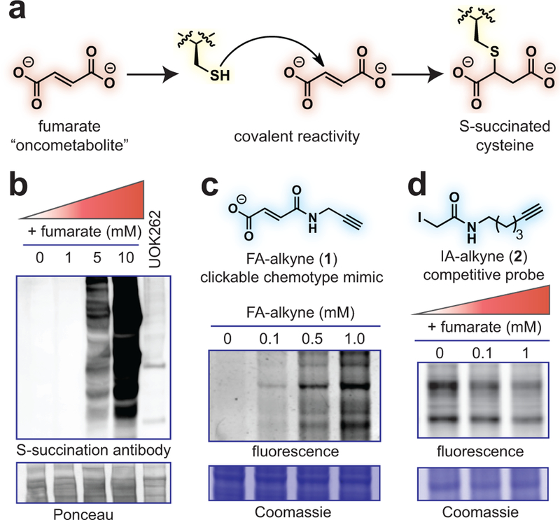
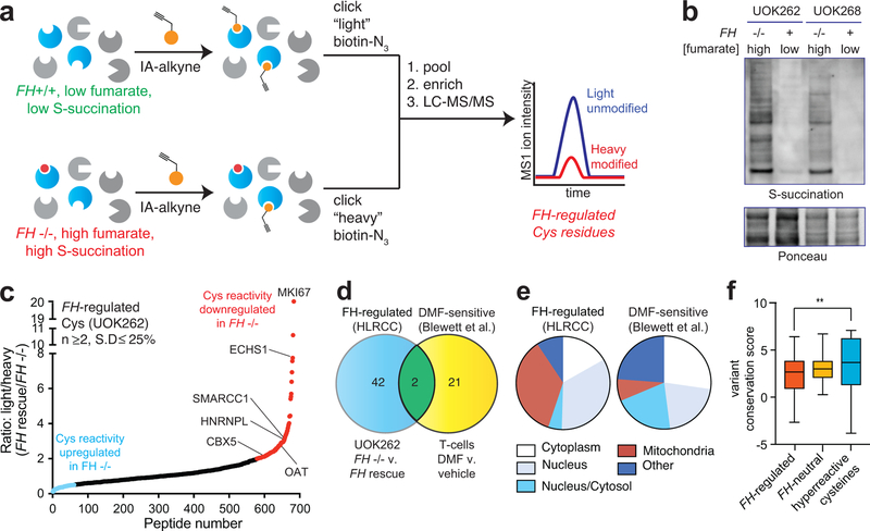
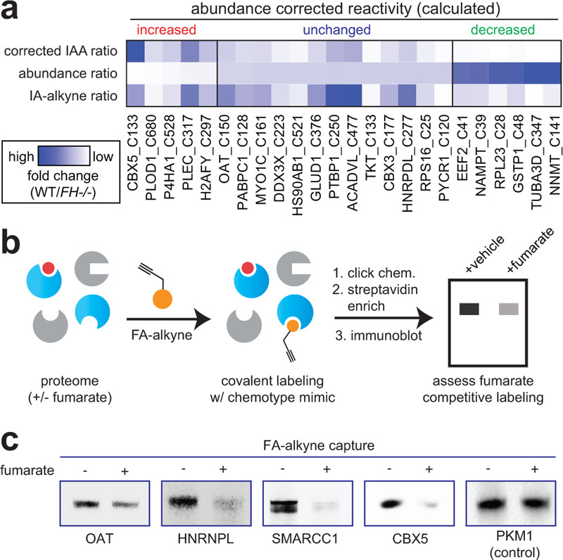
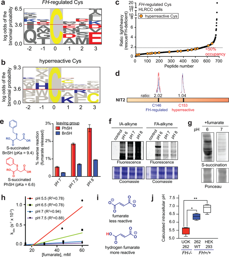
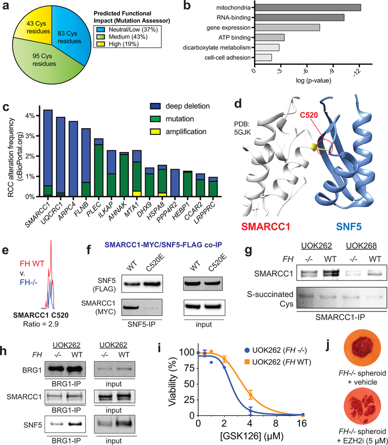
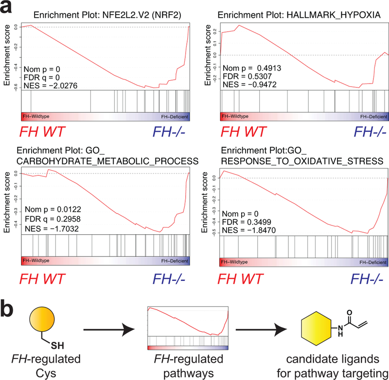
Hereditary cancer disorders often provide an important window into novel mechanisms supporting tumor growth. Understanding these mechanisms thus represents a vital goal. Toward this goal, here we report a chemoproteomic map of fumarate, a covalent oncometabolite whose accumulation marks the genetic cancer syndrome hereditary leiomyomatosis and renal cell carcinoma (HLRCC). We applied a fumarate-competitive chemoproteomic probe in concert with LC-MS/MS to discover new cysteines sensitive to fumarate hydratase (FH) mutation in HLRCC cell models. Analysis of this dataset revealed an unexpected influence of local environment and pH on fumarate reactivity, and enabled the characterization of a novel FH-regulated cysteine residue that lies at a key protein-protein interface in the SWI-SNF tumor-suppressor complex. Our studies provide a powerful resource for understanding the covalent imprint of fumarate on the proteome and lay the foundation for future efforts to exploit this distinct aspect of oncometabolism for cancer diagnosis and therapy.


Conflict of interest statement

Competing interests

The authors declare no competing financial interests.

Figures

Figure 1.
Figure 1.
Fumarate is a covalent oncometabolite. (a) Covalent labeling of cysteine residues by fumarate yields the PTM S-succination. (b) Applying S-succinated Cys immunoblotting to establish the concentration of fumarate required for covalent protein labeling. HEK-293 proteomes were treated with fumarate (0, 1, 5, 10 mM) for 15 h prior to western blotting. (c) Applying fumarate alkyne (FA-alkyne, 1) to visualize reactivity of the fumarate chemotype. HEK-293 proteomes were treated with FA-alkyne (0, 0.1, 0.5, 1 mM) for 15 h prior to click chemistry and SDS-PAGE. (d) Applying iodoacetamide alkyne (IA-alkyne, 2) as a competitive probe of covalent fumarate labeling. HEK-293 proteomes were incubated with fumarate for 15 h prior to treatment with 100 μM IA-alkyne for 1 h followed by desalting, click chemistry, and SDS-PAGE. Representative images from two independent experiments are shown in b-d. Uncropped scans of gels and immunoblots are provided in Supplementary Fig. 10.
Figure 2.
Figure 2.
Global chemoproteomic profiling of FH-regulated cysteine residues. (a) Applying a competitive chemoproteomic platform to study the oncometabolite fumarate. Comparison of proteomes from FH−/− (UOK262) and FH+/+ (UOK262WT) cells are used to define “FH-regulated” Cys residues. (b) S-succination in HLRCC cells is dependent on FH mutation. Representative image from two independent experiments is shown. Uncropped scans of immunoblot is provided in Supplementary Fig. 10. (c) FH-regulated Cys residues identified in UOK262 cells (n ≥2, SD ≤25%). (d) Overlap of FH-regulated Cys residues (R ≥2, n ≥1) with DMF-regulated Cys residues identified in Blewett et al (R ≥2, n≥1). (e) Subcellular localization of FH-regulated and DMF-sensitive Cys residues. (f) Conservation of FH-regulated, FH-neutral, and hyperreactive cysteine residues identified in Weerapana et al. Peptides with the highest R values from each dataset (n=50) were used for analysis. Data is presented as box and whiskers plot with box representing 25th-75th percentile, horizontal line representing median, and whiskers representing minimum and maximum values. Statistical significance was assessed using Student’s t-test (two-tailed, unpaired); **P < 0.01. Data for individual proteins is available in supplementary datasets and can be searched via a web interface at ccr2.cancer.gov/resources/Cbl/proteomics/fumarate.
Figure 3.
Figure 3.
Analyzing the reactivity and abundance of FH-regulated cysteines. (a) Heat map illustrating strategy for correcting Cys reactivity ratios measured in UOK262 (FH-deficient) and UOK262WT (FH rescue) cells using whole proteome MudPIT LC-MS/MS data. Adjusting for protein abundance can lead to an increase in calculated reactivity (left protein subset, red), an insignificant change (middle protein subset, blue), or a decrease in calculated reactivity (right protein subset, green). An extended graphic of protein families harboring high confidence, abundance-corrected FH-regulated cysteine residues is provide in Supplementary Fig. 2d. (b) Validating FH-regulated Cys residues using the clickable chemotype mimic FA-alkyne. (c) FA-alkyne capture of proteins that contain FH-regulated Cys residues is competed by fumarate (3 h pre-incubation with 1 mM fumarate; then 15 h treatment with 100 μM FA-alkyne). Representative images from two independent experiments are shown. Uncropped scans of immunoblots are provided in Supplementary Fig. 10.
Figure 4.
Figure 4.
Establishing the molecular determinants of fumarate-protein interactions. (a) Motif analysis of FH-regulated Cys residues. 50 cysteines found to be most FH-regulated in this study (highest R values, n≥2, SD≤25%) were used for the analysis. (b) Motif analysis of hyperreactive Cys residues identified in Weerapana et al. 50 cysteines found to be most hyperreactive were used for the analysis. (c) FH-regulated Cys residues are anti-correlated with Cys-reactivity. (d) FH mutation preferentially modulates the reactivity of a distal Cys in NIT2 (n = 3 independent experiments). (e) The reversibility of S-succinated model thiols is slow and dependent on leaving group pKa.. S-succinated thiols (1 mM) were incubated in 100 mM Tris at 37 °C for 24 h, prior to quantification of DMF release by fluorescence assay. Data is presented as mean ± s.e.m.; n = 3/group. (f) Influence of pH on IA-alkyne and FA-alkyne reactivity. Left: IA-alkyne (1h; 100 μM); Right: FA-alkyne (15 h, 1 mM). (g) Influence of pH on proteomic S-succination (16h; 5mM). Representative images from two independent experiments are shown in f-g. Uncropped scans of gels and immunoblots are provided in Supplementary Fig. 10. (h) pH-dependent reaction kinetics of fumarate with thiophenol (n = 3 independent experiments). R2 = goodness of fit. (i) Structures of fumarate and hydrogen fumarate. (j) FH−/− cell lines exhibit decreased intracellular pH relative to FH+/+ lines. pH values were determined using pHrodo Green Statistical significance was assessed using Student’s t-test (two-tailed, unpaired); n = 4/group, *P < 0.05, **P < 0.01.
Figure 5.
Figure 5.
Functional analyses of FH-regulated Cys residues. (a) Percentage of FH-regulated Cys residues predicted to be functional using the informatics tool Mutation Assessor (R ≥1.5, n ≥2). (b) Gene ontology analysis of FH-regulated Cys residues. (c) Correlation between genes containing FH-regulated Cys residues and genetic alterations in kidney cancer (RCC). Statistical significance was assessed using Student’s t-test (two-tailed, unpaired); **P < 0.01. (d) SMARCC1 C520 lies in the SWIRM domain and is highly conserved. (e) SMARCC1 C520 lies at the SNF5 subunit interface. (f) SMARCC1 C520 undergoes FH-regulated changes in occupancy in HLRCC cells (n = 3 independent experiments). (g) SMARCC1 C520E mutation limits co-immunoprecipitation with SNF5 in HEK-293 cells co-overexpressing FLAG-tagged SNF5 with Myc-tagged SMARCC1. (h) SMARCC1 S-succination can be detected in FH-deficient and FH WT HLRCC cell lines after IP of endogenous SMARCC1. (i) SNF5 demonstrates decreased co-immunoprecipitation and decreased levels in FH−/− HLRCC cells. Left: Results from SWI/SNF complex co-immunoprecipitation with BRG1 antibody. Right: Endogenous levels of SWI/SNF complex members in HLRCC cells. Representative images from two independent experiments are shown in g-i. Uncropped scans of immunoblots are provided in Supplementary Fig. 10. (j) EZH2 inhibitors exhibit modest selectivity for FH-deficient HLRCC cells. UOK262 FH −/− or FH WT spheroids were treated with GSK126 (21 days; 1, 2, 4, 8, 16 μM) and % viability plotted relative to the vehicle treated spheroids. Data is presented as mean ± s.d.; n = 3/group. (k) EZH2 inhibitors are toxic to HLRCC spheroids. UOK262 FH −/− spheroids were treated with vehicle or EPZ6438 (14 days; 5 μM). Figure is representative of 6 replicates.
Figure 6.
Figure 6.
(a) Gene set enrichment analysis (GSEA) of FH-regulated Cys residues highlights pathways whose activity is functionally upregulated in HLRCC cells. (b) Concept of applying chemoproteomic pathway discovery data to identify new candidate ligands for pathway targeting.

References

    1. Launonen V et al. Inherited susceptibility to uterine leiomyomas and renal cell cancer. Proc Natl Acad Sci U S A 98, 3387–3392, 10.1073/pnas.051633798 (2001). - DOI - PMC - PubMed
    1. Tomlinson IP et al. Germline mutations in FH predispose to dominantly inherited uterine fibroids, skin leiomyomata and papillary renal cell cancer. Nat Genet 30, 406–410, 10.1038/ng849 (2002). - DOI - PubMed
    1. Meier JL Metabolic mechanisms of epigenetic regulation. ACS Chem Biol 8, 2607–2621, 10.1021/cb400689r (2013). - DOI - PMC - PubMed
    1. Isaacs JS et al. HIF overexpression correlates with biallelic loss of fumarate hydratase in renal cancer: novel role of fumarate in regulation of HIF stability. Cancer Cell 8, 143–153, 10.1016/j.ccr.2005.06.017 (2005). - DOI - PubMed
    1. Pollard PJ et al. Accumulation of Krebs cycle intermediates and over-expression of HIF1alpha in tumours which result from germline FH and SDH mutations. Hum Mol Genet 14, 2231–2239, 10.1093/hmg/ddi227 (2005). - DOI - PubMed

Publication types

MeSH terms

Supplementary concepts