

New Light Shed On The Enigmatic Eosinophil Granulocyte; A Versatile Cell Of The Immune System
- PMID: 30720250
- PMCID: PMC6357243
New Light Shed On The Enigmatic Eosinophil Granulocyte; A Versatile Cell Of The Immune System
Abstract
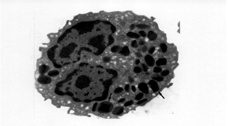
Eosinophils normally constitute only a few per cent of circulating leukocytes, though they are more numerous in tissues vulnerable to attack by environmental microorganisms. Eosinophils can kill invasive parasites, but they also have immunoregulatory functions and may be involved in, for example, the connective tissue remodeling that occurs in conjunction with inflammation. Although their effects may be beneficial to the host, for instance in the event of helminthic infestation, they may also cause tissue damage, for example in allergy and asthma. Recent years have witnessed significant advances in our knowledge of these fascinating but still enigmatic cells. The eosinophil granulocyte was first described in 1879 by Paul Ehrlich, who discovered a blood cell that had high affinity for acid dyes and, in particular, eosin (1) (Figure 1). Eosin, which gives the cells their characteristic red-orange color, is named after Eos, the goddess of dawn in Greek mythology. Paul Ehrlich also put forward the hypothesis that eosinophils develop in the bone marrow (2) and exert their functions in the peripheral tissues. All vertebrates seem to have eosinophils in their blood, but these cells have also been described in more primitive organisms such as sharks, turtles and snakes (3). However. there are differences in morphology, and a comparison of eosinophils from different species also reveals considerable differences in the protein content of their characteristic cytoplasmic granules. For example, in contrast to many vertebrates, eosinophils from the cat, rhino, hyena and okapi lack peroxidase (3).
Figures


Tissue cells, e.g. lymphocytes (Ly) or macrophages (Mø), stimulate endothelial cells in postcapillary venules to express surface adhesion molecules on their surface.
Initially the cell is ”marginated” in the blood vessel and starts ”rolling” along the endothelium, a process mediated by adhesion molecules of the selectin type.
The eosinophil granulocytes are activated, in particular by inflammatory mediators released by the endothelial cells, and subsequently bind strongly to the endothelial cells. This firm binding is mediated through ß1- and ß2-integrins. The activation of ß2-integrins also cause a shape change and the eosinophil becomes flattened.
Several inflammatory mediators produced at the site of inflammation stimulate the eosinophil granulocytes to leave the blood vessel, using its ß1- and ß2-integrins, and enter the tissue where it exerts its function.

The cell phagocytoses bacteria with immunoglobulins and complement molecules on their surface. The bacteria are enclosed in an invagination of cell membrane that ñnally becomes a phagosome of their cytoplasm. Specific granules are transported to the phagosome (1), and after fusion (2) the granular content is released (3) and the cytotoxic process started.
The eosinophil granulocyte attacks a parasite too big to phagocytose, resulting in ”frustrated” phagocytosis. The granules are transported to the part of the cell membrane that is in contact with the parasite (1). The membranes of the granule and the eosinophil plasma membrane fuse (2) followed by release of the granule content onto the surface of the parasite (3) where the cytotoxic effects are exerted. (artist: Piroska von Gegerfeldt)
Similar articles
-
[New light shed on the enigmatic eosinophilic granulocyte. Both a friend and an enemy].Lakartidningen. 1998 Feb 25;95(9):850-6, 859. Lakartidningen. 1998. PMID: 9531752 Review. Swedish.
-
The early history of the eosinophil.Clin Exp Allergy. 2015 Mar;45(3):575-82. doi: 10.1111/cea.12480. Clin Exp Allergy. 2015. PMID: 25544991 Review.
-
Eosinophils: multifunctional and distinctive properties.Int Arch Allergy Immunol. 2013;161 Suppl 2(0 2):3-9. doi: 10.1159/000350662. Epub 2013 May 29. Int Arch Allergy Immunol. 2013. PMID: 23711847 Free PMC article. Review.
-
Eosinophils: a review.Vet Res Commun. 1992;16(1):11-44. doi: 10.1007/BF01839203. Vet Res Commun. 1992. PMID: 1598753 Review.
-
Circulating eosinophils in asthma, allergic rhinitis, and atopic dermatitis lack morphological signs of degranulation.Clin Exp Allergy. 2005 Oct;35(10):1334-40. doi: 10.1111/j.1365-2222.2005.02335.x. Clin Exp Allergy. 2005. PMID: 16238793
Cited by
-
Hemogram study of an artificially feeding tree shrew (Tupaia belangeri chinensis).Exp Anim. 2020 Jan 29;69(1):80-91. doi: 10.1538/expanim.19-0079. Epub 2019 Sep 17. Exp Anim. 2020. PMID: 31527336 Free PMC article.
References
-
- Ehrlich P. Ueber die specifischen granulationen des Blutes. Arch Anat Physiol Lpz. 1879; 3 Physiol Abt: 571-579.
-
- Ehrlich P. Beitrage zur kenntniss der granulierten bindergewebszellen und der eosinophilen leukocyten. Arch Anat Physiol Lpz. 1879; 3 Physiol Abt: 166-169.
-
- Spry CJF. Eosinophils. A comprehensive review and guide to the scientific and medical literature. Oxford: Oxford University Press, 1988.
-
- Sanderson CJ. Interleukin-5, eosinophils, and disease. Blood 1992; 79: 3101-3109. - PubMed
-
- Lopez AF, Williamson DJ, Gamble JR, Begley CG, Harlan JM, Klebanoff SJ, et al. Recombinant human granulocyte-macrophage colony stimulating factor stimulates in vitro mature human neutrophil and eosinophil function, surface receptor expression, and survival. J Clin Invest 1986; 78: 1220-1228. - PMC - PubMed
Publication types
LinkOut - more resources
Full Text Sources
Miscellaneous