Comparative Study
. 2019 May;43(5):695-702.
doi: 10.1097/PAS.0000000000001227.

Expanding the Spectrum of Intraosseous Rhabdomyosarcoma: Correlation Between 2 Distinct Gene Fusions and Phenotype

Affiliations
Comparative Study

Expanding the Spectrum of Intraosseous Rhabdomyosarcoma: Correlation Between 2 Distinct Gene Fusions and Phenotype

Narasimhan P Agaram et al. Am J Surg Pathol. 2019 May.

Abstract

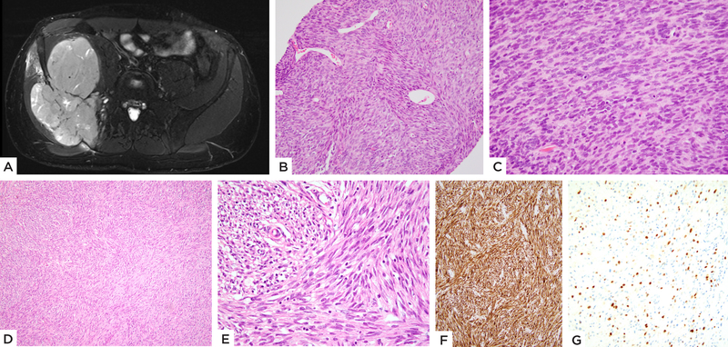
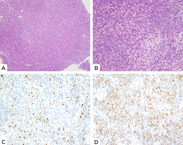
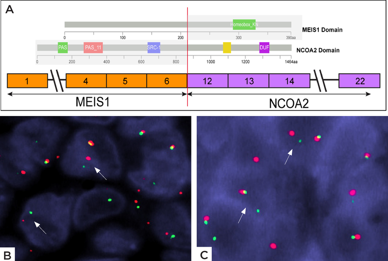
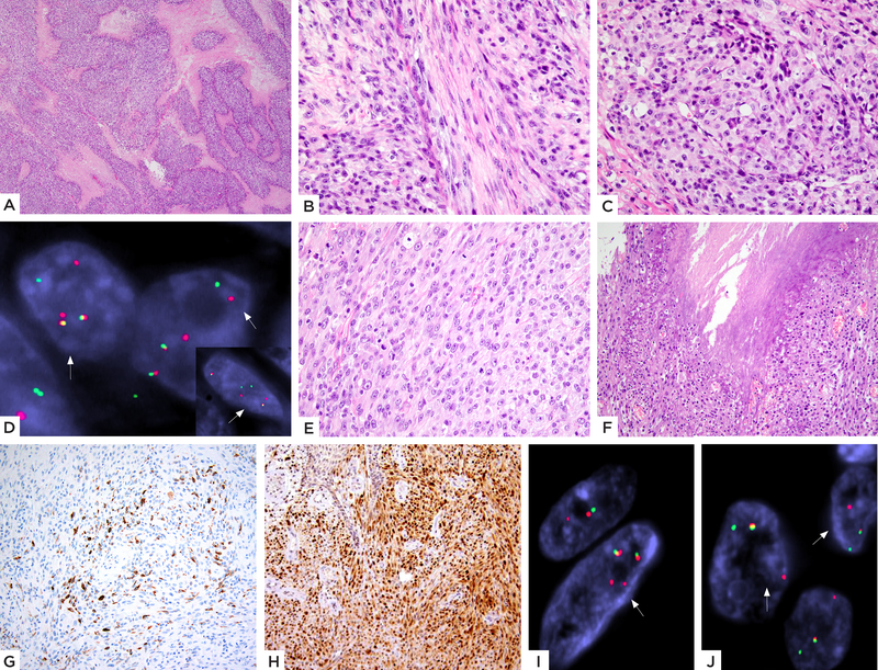
Primary intraosseous rhabdomyosarcomas (RMSs) are extremely rare. Recently 2 studies reported 4 cases of primary intraosseous RMS with EWSR1/FUS-TFCP2 gene fusions, associated with somewhat conflicting histologic features, ranging from spindle to epithelioid. In this study we sought to further investigate the pathologic and molecular abnormalities of a larger group of intraosseous RMSs by a combined approach using targeted RNA sequencing analysis and fluorescence in situ hybridization (FISH). We identified 7 cases, 3 males and 4 females, all in young adults, age range 20 to 39 years (median, 27 y). Three cases involved the pelvis, 2 involved the femur and 1 each involved the maxilla and the skull. Molecular studies identified recurrent gene fusions in all 7 cases tested, including: a novel MEIS1-NCOA2 fusion in 2 cases, EWSR1-TFCP2 in 3 cases, and FUS-TFCP2 gene fusions in 1 case. One case showed a FUS gene rearrangement, without a TFCP2 gene abnormality by FISH. The MEIS1-NCOA2-positive cases were characterized by a more primitive and fascicular spindle cell appearance, while the EWSR1/FUS rearranged tumors had a hybrid spindle and epithelioid phenotype, with more abundant eosinophilic cytoplasm and mild nuclear pleomorphism. Immunohistochemically, all tumors were positive for desmin and myogenin (focal). In addition, 4 tumors with TFCP2-associated gene fusions also coexpressed ALK and cytokeratin. In conclusion, our results suggest a high incidence of gene fusions in primary RMSs of bone, with 2 molecular subsets emerging, defined by either MEIS1-NCOA2 or EWSR1/FUS-TFCP2 fusions, showing distinct morphology and immunophenotype. Additional studies with larger numbers of cases and longer follow-up data are required to definitively evaluate the biological behavior of these tumors and to establish their relationship to other spindle cell RMS genetic groups.


Figures

Figure 1:
Figure 1:. MEIS1-NCOA2 positive iliac bone rhabdomyosarcoma
(case 1, 22/M). A) MRI showing a large tumor centered in the iliac bone with extraosseous extension. B) Pre-therapy biopsy shows a highly cellular spindle cell neoplasm with scattered branching vessels and insignificant stromal component; C) higher power with monomorphic spindle cells arranged in a streaming pattern. D, E) Resected tumor s/p chemotherapy reveals spindle cells in intersecting fascicles and scant eosinophilic cytoplasm. Immunohistochemical stains show F) diffuse positivity for desmin and G) focal Myogenin staining.
Figure 2:
Figure 2:. MEIS1-NCOA2 positive iliac bone rhabdomyosarcoma
(case 2, 39/M). A) Low power showing a densely cellular primitive neoplasm. B) High power reveals round to ovoid and short spindle cells arranged in vague micronodules or whorling patterns. Immunohistochemical stains show C) scattered multifocal Myogenin positivity and D) relatively diffuse MyoD1 staining.
Figure 3:
Figure 3:. Schematic diagram of MEIS1-NCOA2 fusion.
A) ARCHER results revealed a fusion transcript composed of MEIS1 exon 6 fused to NCOA2 exon 12. The proteins domains of MEIS1 and NCOA2 are illustrated above. B) FISH studies showing break-apart signals of B) NCOA2 gene and C) MEIS1 gene (red, centromeric; green telomeric).
Figure 4:
Figure 4:. EWSR1-TFCP2 fusion positive rhabdomyosarcoma with a mixed spindle and epithelioid histology.
(A-D) Case 3 (27/F, skull): A) Low power showing a cellular neoplasm with geographic areas of necrosis. High power showing B) spindle cells with pale eosinophilic cytoplasm arranged in intersecting fascicles at 90-degrees and C) areas with distinct epithelioid features. D) FISH studies show break-apart signals for both TFCP2 and EWSR1 (inset) genes (red, centromeric, green, telomeric). (E-J) Case 4 (33/F, maxilla): E) Cellular neoplasm composed of plump ovoid to short spindle cells with moderately abundant pale eosinophilic cytoplasm, arranged in solid sheets or streaming pattern and F) areas of necrosis. Immunohistochemical stains show G) patchy positivity for desmin and H) relatively diffuse nuclear staining for MyoD1. FISH analysis showed the presence of break-apart split signals for both I) EWSR1 and J) TFCP2 genes (red, centromeric; green, telomeric)
Figure 5:
Figure 5:. TFCP2 fusion positive subset showing mainly spindle cell morphology.
(A-C) EWSR1-TFCP2 fusion positive femur rhabdomyosarcoma (case 5, 20/M). A) Cellular neoplasm involving the bone and completely replacing marrow spaces with spindle cells arranged in intersecting fascicles or vague storiform, with B) pale eosinophilic cytoplasm and ovoid to fusiform nuclei with high mitotic activity. C) Immunohistochemical stain shows diffuse MYOD1 positivity. (D-F) FUS-TFCP2 positive iliac bone tumor (case 6, 37/F). D) Low power showing a cellular neoplasm diffusely involving bone. E) High power reveals short spindle cells arranged in short fascicles. F) Immunohistochemical stain shows focal Myogenin positivity (Inset: diffuse ALK immunoreactivity).

References

    1. Andrade CR, Trento GDS, Jeremias F, et al. Rabdomyosarcoma of the Mandible: An Uncommon Clinical Presentation. J Craniofac Surg. 2018;29:e221–e224. - PubMed
    1. Cemiloglu R, Tekalan SA, Patiroglu T, et al. Rhabdomyosarcoma of the temporal bone: clinical report. Arch Otorhinolaryngol. 1987;244:195–197. - PubMed
    1. Chasin WD. Rhabdomyosarcoma of the temporal bone. Ann Otol Rhinol Laryngol Suppl. 1984;112:71–73. - PubMed
    1. de Melo ACR, Lyra TC, Ribeiro ILA, et al. Embryonal rhabdomyosarcoma in the maxillary sinus with orbital involvement in a pediatric patient: Case report. World J Clin Cases. 2017;5:440–445. - PMC - PubMed
    1. Iatrou I, Theologie-Lygidakis N, Schoinohoriti O, et al. Rhabdomyosarcoma of the maxillofacial region in children and adolescents: Report of 9 cases and literature review. J Craniomaxillofac Surg. 2017;45:831–838. - PubMed

Publication types

MeSH terms