

Resting state connectivity differences in eyes open versus eyes closed conditions
- PMID: 30720907
- PMCID: PMC6865559
- DOI: 10.1002/hbm.24539
Resting state connectivity differences in eyes open versus eyes closed conditions
Abstract
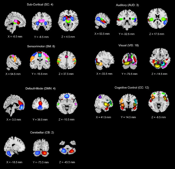
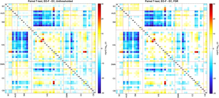
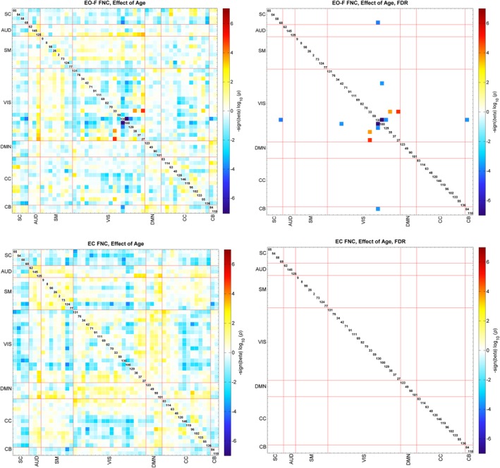
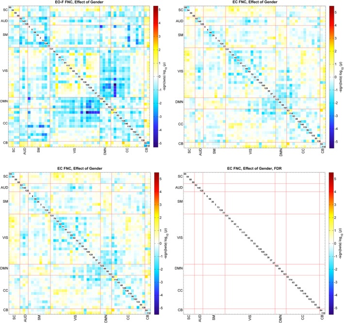
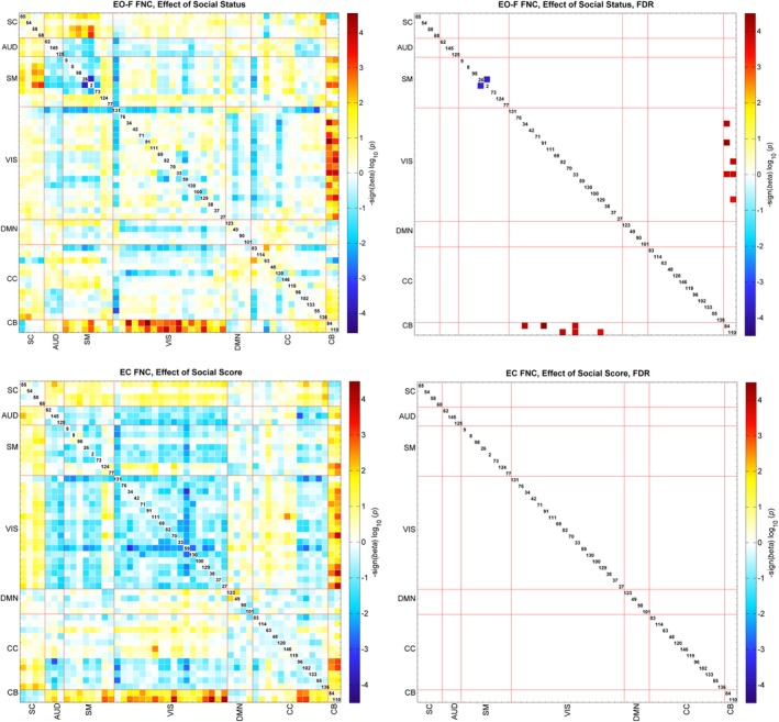
Functional magnetic resonance imaging data are commonly collected during the resting state. Resting state functional magnetic resonance imaging (rs-fMRI) is very practical and applicable for a wide range of study populations. Rs-fMRI is usually collected in at least one of three different conditions/tasks, eyes closed (EC), eyes open (EO), or eyes fixated on an object (EO-F). Several studies have shown that there are significant condition-related differences in the acquired data. In this study, we compared the functional network connectivity (FNC) differences assessed via group independent component analysis on a large rs-fMRI dataset collected in both EC and EO-F conditions, and also investigated the effect of covariates (e.g., age, gender, and social status score). Our results indicated that task condition significantly affected a wide range of networks; connectivity of visual networks to themselves and other networks was increased during EO-F, while EC was associated with increased connectivity of auditory and sensorimotor networks to other networks. In addition, the association of FNC with age, gender, and social status was observed to be significant only in the EO-F condition (though limited as well). However, statistical analysis did not reveal any significant effect of interaction between eyes status and covariates. These results indicate that resting-state condition is an important variable that may limit the generalizability of clinical findings using rs-fMRI.
Keywords: eyes closed; eyes open; functional network connectivity; independent component analysis; resting state fMRI.
© 2019 Wiley Periodicals, Inc.
Figures

References
-
- Agcaoglu, O. , Miller, R. , Damaraju, E. , Rashid, B. , Bustillo, J. , Cetin, M. S. , … Calhoun, V. D. (2018). Decreased hemispheric connectivity and decreased intra‐ and inter‐ hemisphere asymmetry of resting state functional network connectivity in schizophrenia. Brain Imaging and Behavior, 12(3), 615–630. 10.1007/s11682-017-9718-7 - DOI - PMC - PubMed
-
- Agcaoglu, O. , Miller, R. , Mayer, A. R. , Hugdahl, K. , & Calhoun, V. D. (2016). Increased spatial granularity of left brain activation and unique age/gender signatures: A 4D frequency domain approach to cerebral lateralization at rest. Brain Imaging and Behavior, 10(4), 1004–1014. 10.1007/s11682-015-9463-8 - DOI - PMC - PubMed
Publication types
MeSH terms
Grants and funding
LinkOut - more resources
Full Text Sources
