. 2019 May;37(5):652-662.
doi: 10.1002/stem.2988. Epub 2019 Feb 22.

Extracellular Vesicle-Educated Macrophages Promote Early Achilles Tendon Healing

Affiliations

Extracellular Vesicle-Educated Macrophages Promote Early Achilles Tendon Healing

Connie S Chamberlain et al. Stem Cells. 2019 May.

Abstract

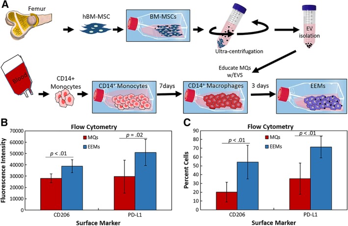
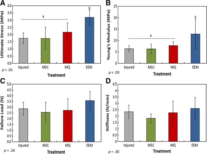
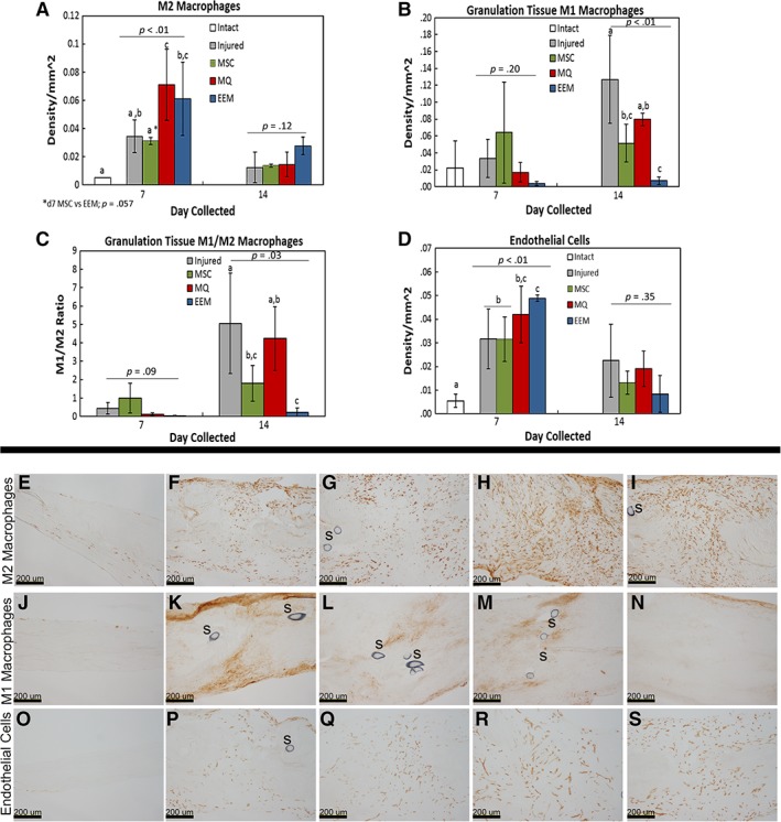
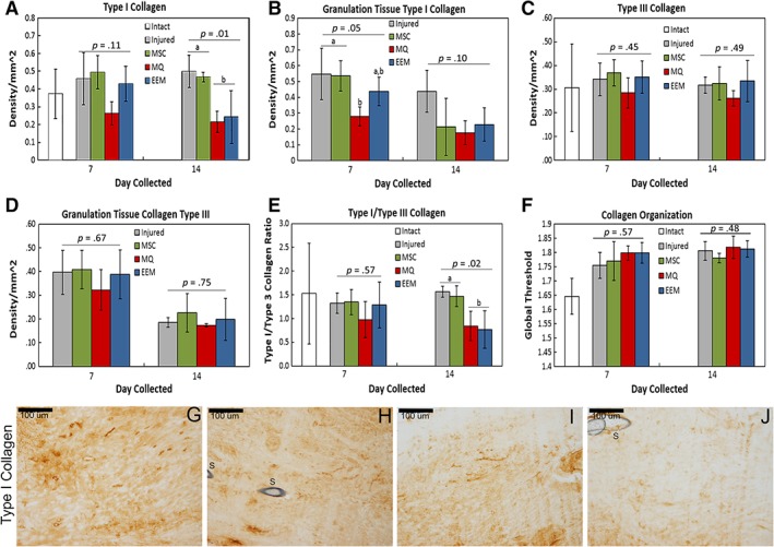
Tendon healing follows a complex series of coordinated events, which ultimately produces a mechanically inferior tissue more scar-like than native tendon. More regenerative healing occurs when anti-inflammatory M2 macrophages play a more dominant role. Mesenchymal stromal/stem cells (MSCs) are able to polarize macrophages to an M2 immunophenotype via paracrine mechanisms. We previously reported that coculture of CD14+ macrophages (MQs) with MSCs resulted in a unique M2-like macrophage. More recently, we generated M2-like macrophages using only extracellular vesicles (EVs) isolated from MSCs creating "EV-educated macrophages" (also called exosome-educated macrophages [EEMs]), thereby foregoing direct use of MSCs. For the current study, we hypothesized that cell therapy with EEMs would improve in vivo tendon healing by modulating tissue inflammation and endogenous macrophage immunophenotypes. We evaluated effects of EEMs using a mouse Achilles tendon rupture model and compared results to normal tendon healing (without any biologic intervention), MSCs, MQs, or EVs. We found that exogenous administration of EEMs directly into the wound promoted a healing response that was significantly more functional and more regenerative. Injured tendons treated with exogenous EEMs exhibited (a) improved mechanical properties, (b) reduced inflammation, and (c) earlier angiogenesis. Treatment with MSC-derived EVs alone were less effective functionally but stimulated a biological response as evidenced by an increased number of endothelial cells and decreased M1/M2 ratio. Because of their regenerative and immunomodulatory effects, EEM treament could provide a novel strategy to promote wound healing in this and various other musculoskeletal injuries or pathologies where inflammation and inadequate healing is problematic. Stem Cells 2019;37:652-662.

Keywords: Achilles tendon injury; Extracellular vesicles; Macrophages; Mesenchymal stromal cells; Tendon healing.


Conflict of interest statement

The authors report no potential conflicts of interest.

Figures

Figure 1
Figure 1
Fabrication and characterization of exosome‐educated macrophages (EEMs). (A): Bone marrow mesenchymal stromal/stem cells (MSCs) were culture expanded. Extracellular vesicles (EVs) were then isolated from the MSCs via ultracentrifugation. CD14+ monocytes were obtained from human peripheral blood. Monocytes were cultured for 7 days, activated to CD14+ macrophages, and then educated with EVs for 3 days, producing EEMs. (B, C): CD14+ macrophages (control macrophages [MQs]) and EEMs were measured for alternatively activated macrophage cell surface markers by flow cytometry. (B): Fluorescence intensity and (C) percent cells indicate more EEMs were positive for CD206, and PD‐L1 compared with the MQs. *, p values are results of Fisher's least significant difference (LSD) post hoc pairwise analysis (p < .05). Values are expressed as mean cell numbers ± SD.
Figure 2
Figure 2
Mechanical results of the healing Achilles tendon after exosome‐educated macrophage (EEM) treatment. Data indicate (A) ultimate stress, (B) Young's modulus, (C) failure load, and (D) stiffness of the Achilles tendons 14 days after injury after treatments with Hank's balanced saline solution (“injured”), mesenchymal stromal/stem cells, control macrophages, or EEMs. EEM treatment significantly improved (A) ultimate stress and (B) Young's modulus compared with all other treatments. No significance was found between (C) failure load and (D) stiffness. Bars without a common superscript within a graph indicate they are significantly different (results of Fisher's LSD post hoc pairwise analysis, p < .05). Results are expressed as mean ± SD.
Figure 3
Figure 3
Immunohistochemistry results of the day 7 and 14 healing tendon. Graphs of (A) total M2 macrophages, (B) M1 macrophages localized to the granulation tissue, (C) M1/M2 macrophage ratio, and (D) endothelial cells within the healing Achilles tendon after treatment with Hank's balanced saline solution (injured), mesenchymal stromal/stem cells (MSCs), control macrophages (MQs), or exosome‐educated macrophages (EEMs). Representative images of the (E–I) M2 macrophages, (J–N) M1 macrophages, (O–S) endothelial cells by the (E, J, O) intact, (F, P) day 7 injured, (G, Q) MSCs, (H, R) MQs, (I, S) EEM, and (K) day 14 injured, (L) MSC, (M) MQ, and (N) EEM. “S” indicates sutures within the tissue. Data are considered significantly different (p < .05) based on Fisher's LSD post hoc pairwise analysis. Within each bar graph, samples without a common superscript (a, b, c) are deemed significantly different. Values are expressed as mean cell numbers ± SD.
Figure 4
Figure 4
Immunohistochemistry results of extracellular matrix factors by day 7 and 14 healing tendon. Immunohistochemistry results showing the effects of mesenchymal stromal/stem cells (MSCs), control macrophages (MQs), and exosome‐educated macrophages (EEMs) on (A) type I collagen throughout entire tendon, (B) type I collagen within the granulation tissue, (C) type III collagen throughout entire tendon, (D) type III collagen within the granulation tissue, (E) type I/type III collagen ratio, and (F) collagen organization. (G–J): Representative images of type I collagen by the day 14 Achilles tendon after (G) injured (control), (H) MSC, (I) MQ, or (J) EEM treatment. Data are considered significantly different (p < .05) based on Fisher's LSD post hoc pairwise analysis. Within each bar graph, samples without a common superscript (a, b) are deemed significantly different. Values are expressed as mean cell numbers ± SD.

References

    1. Mantovani A, Biswas SK, Galdiero MR et al. Macrophage plasticity and polarization in tissue repair and remodelling. J Pathol 2013;229:176–185. - PubMed
    1. Murray PJ. Macrophage polarization. Annu Rev Physiol 2017;79:541–566. - PubMed
    1. Martinez FO, Helming L, Gordon S. Alternative activation of macrophages: An immunologic functional perspective. Annu Rev Immunol 2009;27:451–483. - PubMed
    1. Godwin JW, Pinto AR, Rosenthal NA. Macrophages are required for adult salamander limb regeneration. Proc Natl Acad Sci USA 2013;110:9415–9420. - PMC - PubMed
    1. Sindrilaru A, Peters T, Wieschalka S et al. An unrestrained proinflammatory M1 macrophage population induced by iron impairs wound healing in humans and mice. J Clin Invest 2011;121:985–997. - PMC - PubMed

Publication types

MeSH terms