Review
. 2019 May;156(7):2024-2040.
doi: 10.1053/j.gastro.2019.01.259. Epub 2019 Feb 2.

Early Detection of Pancreatic Cancer: Opportunities and Challenges

Affiliations
Review

Early Detection of Pancreatic Cancer: Opportunities and Challenges

Aatur D Singhi et al. Gastroenterology. 2019 May.

Abstract

Most patients with pancreatic ductal adenocarcinoma (PDAC) present with symptomatic, surgically unresectable disease. Although the goal of early detection of PDAC is laudable and likely to result in significant improvement in overall survival, the relatively low prevalence of PDAC renders general population screening infeasible. The challenges of early detection include identification of at-risk individuals in the general population who would benefit from longitudinal surveillance programs and appropriate biomarker and imaging-based modalities used for PDAC surveillance in such cohorts. In recent years, various subgroups at higher-than-average risk for PDAC have been identified, including those with familial risk due to germline mutations, a history of pancreatitis, patients with mucinous pancreatic cysts, and elderly patients with new-onset diabetes. The last 2 categories are discussed at length in terms of the opportunities and challenges they present for PDAC early detection. We also discuss current and emerging imaging modalities that are critical to identifying early, potentially curable PDAC in high-risk cohorts on surveillance.

Keywords: Biomarker; Pre-diagnostic; Risk Stratification.


Conflict of interest statement

Conflicts of interest:

A.D.S. = Foundation Medicine (honorarium)

E.J.K = Philips Healthcare (sponsored research agreement), GE Healthcare (in-kind grant)

Figures

Figure 1:
Figure 1:
The pathology of Intraductal Papillary Mucinous Neoplasms (IPMNs). The macroscopic and microscopic features of IPMNs are typically characterized by involvement of the main pancreatic duct, branch duct (shown here) or both. IPMNs are composed of mucinous epithelium that may be either flat or papillary in appearance. Based on the degree of cytoarchitectural atypia, IPMNs can be classified with low-grade or high-grade dysplasia. The most important prognosticator, however, is the absence or presence of an associated invasive pancreatic ductal adenocarcinoma (PDAC).
Figure 2:
Figure 2:
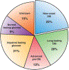
Distribution of glycemic status based on fasting blood glucose levels in a population-based PDAC cohort (N = 219)
Figure 3:
Figure 3:
Common imaging modalities for PDAC including endoscopic ultrasound (EUS, top), computed tomography (CT, middle), and magnetic resonance imaging (MRI, bottom). Each image shows a patient with a ~2 cm lesion in the body of the pancreas. Each modality has advantages and disadvantages for the purposes of early detection of PDAC. A few practical considerations are enumerated.
Figure 4:
Figure 4:
The “future” of PDAC early detection. Currently, the majority of PDAC are diagnosed at a late stage of their natural history, when they are symptomatic, if not surgically unresectable. Individuals with a family history or with cystic lesions represent high-risk cohorts that can be entered into surveillance programs, but only comprise a subset of patients who develop PDAC. Determination of “sporadic risk” will require multiple input parameters (polygenic risk score, BMI, smoking history, other variables), but has the potential to impact the largest subset of individuals in the general population. Surveillance and diagnosis of asymptomatic PDAC in longitudinally monitored high risk cohorts will require biomarkers with exquisite sensitivity and specificity, to avoid the perils of false negatives and overdiagnosis, respectively. Imaging studies, using a bevy of localization modalities discussed in the text, represents the penultimate step before an intervention such as surgery for removing a potentially “curable” early PDAC.

References

    1. Ryan DP, Hong TS, Bardeesy N. Pancreatic adenocarcinoma. N Engl J Med 2014;371:1039–49. - PubMed
    1. Kleeff J, Korc M, Apte M, et al. Pancreatic cancer. Nat Rev Dis Primers 2016;2:16022. - PubMed
    1. Tsuchiya R, Noda T, Harada N, et al. Collective review of small carcinomas of the pancreas. Ann Surg 1986;203:77–81. - PMC - PubMed
    1. Ishikawa O, Ohigashi H, Imaoka S, et al. Minute carcinoma of the pancreas measuring 1 cm or less in diameter--collective review of Japanese case reports. Hepatogastroenterology 1999;46:8–15. - PubMed
    1. Furukawa H, Okada S, Saisho H, et al. Clinicopathologic features of small pancreatic adenocarcinoma. A collective study. Cancer 1996;78:986–90. - PubMed

Publication types

MeSH terms

Substances