. 2019 Feb 5;10(1):e02734-18.
doi: 10.1128/mBio.02734-18.

A Disintegrin and Metalloproteinase 9 Domain (ADAM9) Is a Major Susceptibility Factor in the Early Stages of Encephalomyocarditis Virus Infection

Affiliations

A Disintegrin and Metalloproteinase 9 Domain (ADAM9) Is a Major Susceptibility Factor in the Early Stages of Encephalomyocarditis Virus Infection

Lindsey E Bazzone et al. mBio. .

Abstract

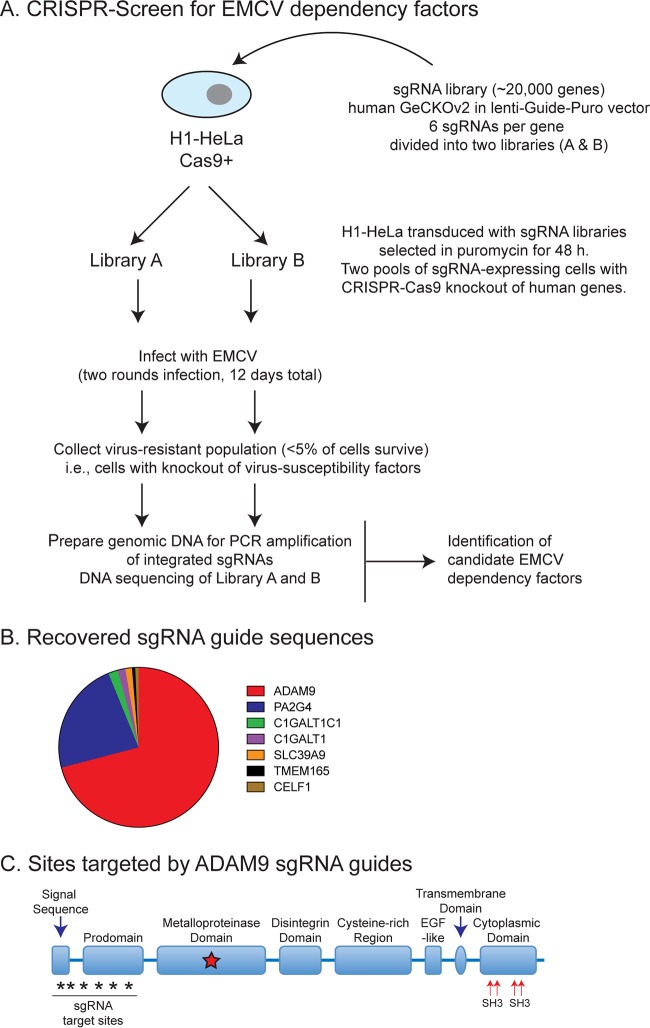
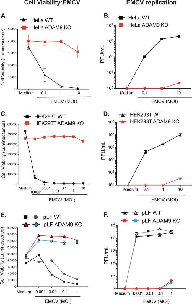
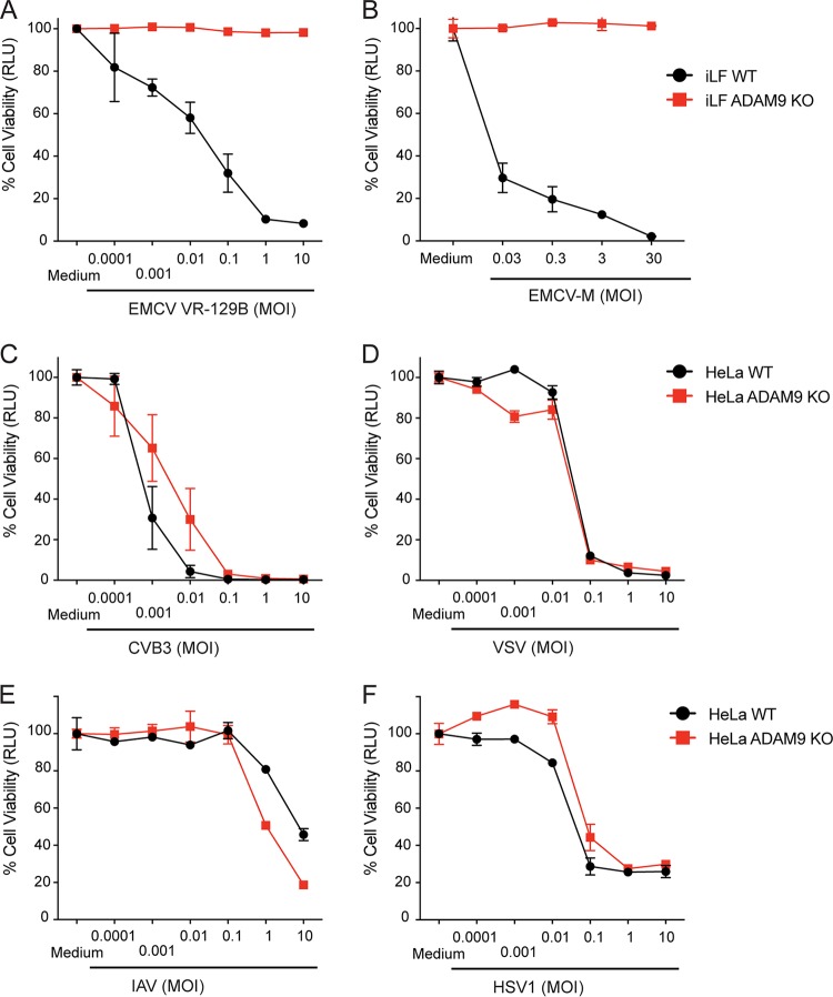
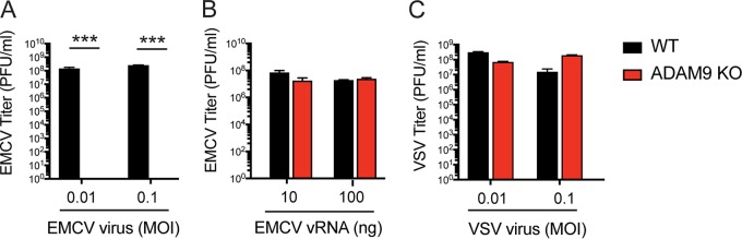
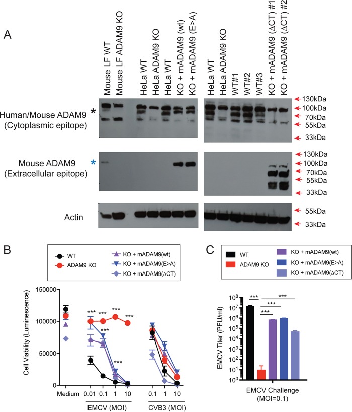
Encephalomyocarditis virus (EMCV) is a picornavirus that produces lytic infections in murine and human cells. Employing a genome-wide CRISPR-Cas9 knockout screen to find host factors required for EMCV infection, we identified a role for ADAM9 in EMCV infection. CRISPR-mediated deletion of ADAM9 in multiple human cell lines rendered the cells highly resistant to EMCV infection and cell death. Primary fibroblasts from ADAM9 KO mice were also strongly resistant to EMCV infection and cell death. In contrast, ADAM9 KO and WT cells were equally susceptible to infection with other viruses, including the picornavirus Coxsackie virus B. ADAM9 KO cells failed to produce viral progeny when incubated with EMCV. However, bypassing EMCV entry into cells through delivery of viral RNA directly to the cytosol yielded infectious EMCV virions from ADAM9 KO cells, suggesting that ADAM9 is not required for EMCV replication post-entry. These findings establish that ADAM9 is required for the early stage of EMCV infection, likely for virus entry or viral genome delivery to the cytosol.IMPORTANCE Viral myocarditis is a leading cause of death in the United States, contributing to numerous unexplained deaths in people ≤35 years old. Enteroviruses contribute to many cases of human myocarditis. Encephalomyocarditis virus (EMCV) infection causes viral myocarditis in rodent models, but its receptor requirements have not been fully identified. CRISPR-Cas9 screens can identify host dependency factors essential for EMCV infection and enhance our understanding of key events that follow viral infection, potentially leading to new strategies for preventing viral myocarditis. Using a CRISPR-Cas9 screen, we identified adisintegrin and metalloproteinase 9 domain (ADAM9) as a major factor required for the early stages of EMCV infection in both human and murine infection.

Keywords: a disintegrin and metalloproteinase 9 domain (ADAM9); encephalomyocarditis virus; functional genomic screen.


Figures

FIG 1
FIG 1
CRISPR-Cas9 knockout screen for EDFs. (A) HeLa cells expressing Cas9 were transduced with the GeCKOv2 sgRNA library, and guide sequences were retrieved from cells that survived multiple rounds of EMCV infection. (B) ADAM9 sgRNA sequences were highly represented in recovered clones; 6 of the 6 sgRNAs targeting ADAM9 were present in recovered clones. GeCKOv2 screen results for genes conferring susceptibility to EMCV-induced cell death. Of the pooled CRISPR-targeted genes, ADAM9- and PA2G4-targeted guide sequences accounted for ∼70% and ∼22%, respectively. (C) Domain structure and sites targeted by sgRNAs.
FIG 2
FIG 2
ADAM9 is a major dependency factor for EMCV infection of human and mouse cells. WT and ADAM9 KO HeLa cells (A and B) and HEK293T cells (C and D) and two independent sets of primary lung fibroblasts (pLF) isolated from WT and ADAM9 KO mice (E and F) were incubated in medium alone or infected with EMCV at various multiplicities of infection (MOI) for 1 h at 37°C, washed to remove free virus, and cultured for 24 h at 37°C. Cell viability (A, C, and E): ATP was quantified in the cell monolayers using CellGlo reagent, revealing that ADAM9 KO cells are highly resistant to EMCV-induced cell death. Virus replication (B, D, and F): EMCV viral titers, measured on BHK21 monolayers and expressed as PFU per ml cell supernatant, showed only small amounts of replicating virus in ADAM9 KO cells. Cell viability data are mean ± SD from 3 replicate wells; plaque assay data are mean ± SD from 2 replicate wells at each dilution. Biological replicate experiments were performed at least twice with similar results.
FIG 3
FIG 3
ADAM9 is required for EMCV infection but does not affect infection by other RNA or DNA viruses. WT and ADAM9 KO cells, including immortalized lung fibroblasts (iLF) and HeLa cells, were challenged with EMCV VR-129B (A), M-variant VR-1479 (B), Coxsackie virus B3 (CVB3) (C), vesicular stomatitis virus (VSV) (D), influenza A virus (IAV) (E), or herpes simplex virus 1 (HSV-1) (F) at various MOIs or incubated in medium alone for 1 h at 37°C. Cell monolayers were washed to remove free virus and incubated for 24 h at 37°C. Viability of infected cells relative to uninfected (medium-only) controls was determined with a CellGlo ATP assay and is expressed in relative luciferase units (RLU). Data are mean ± SD from 3 replicate wells. Biological replicate experiments were performed at least twice with similar results.
FIG 4
FIG 4
ADAM9 is required for EMCV infection but is not directly required for genome replication in the cytosol. WT and ADAM9 KO immortalized lung fibroblasts (iLF) were incubated with EMCV for 1 h at 37°C and washed (A), transfected with EMCV viral RNA (vRNA) in Lipofectamine (B), or incubated with VSV (C). Culture supernatants were harvested after 18 h. Virus replication was measured by quantifying PFU on BHK-21 cells. Data shown are mean ± SD from 3 replicate wells. ***, P < 0.0001, WT versus ADAM9 KO.
FIG 5
FIG 5
Rescue of ADAM9 expression restores EMCV replication in ADAM9 KO cells. WT and ADAM9 KO HeLa cells were transduced with retroviral vectors with wild-type (WT) murine ADAM9 (mADAM9), catalytically inactive mutant ADAM9 (E>A), cytoplasmic-tail-deleted (ΔCT) ADAM9 constructs, or GFP control vectors. (A) WT, KO, and rescue cell lysates were analyzed by Western blot using two different ADAM9 antibodies. Top panel, rabbit anti-human ADAM9 that detects an epitope in the intracellular domain of human ADAM9 and cross-reacts with mouse ADAM9 (black asterisk). Middle panel, goat anti-mouse ADAM9 that detects the extracellular domain of murine ADAM9 but not human ADAM9 (blue asterisk). Bottom panel, anti-actin which detects both human and murine β-actin. Top panel, WT but not KO cells expressed human ADAM9. Middle panel, rescue but not KO cells expressed murine ADAM9. Bottom panel, actin loading control. (B) WT clones, ADAM9 KO clones, and rescued ADAM9-expressing clones were infected with EMCV or CVB3 at various MOIs and incubated at 37°C for 24 h. Viability of EMCV-infected and CVB3-infected clones was measured by CellGlo ATP luminescence. (C) EMCV replication was quantified in infected culture supernatants by plaque assay. Neither the functional sequence of the ADAM9 metalloproteinase domain nor the cytoplasmic tail is required for EMCV infection. ***, P < 0.0001, KO versus WT and KO versus rescue.

Comment in

References

    1. Carocci M, Bakkali-Kassimi L. 2012. The encephalomyocarditis virus. Virulence 3:351–367. doi: 10.4161/viru.20573. - DOI - PMC - PubMed
    1. Helwig FC, Schmidt CH. 1945. A filter-passing agent producing interstitial myocarditis in anthropoid apes and small animals. Science 102:31–33. doi: 10.1126/science.102.2637.31. - DOI - PubMed
    1. Murnane TG, Craighead JE, Mondragon H, Shelokov A. 1960. Fatal disease of swine due to encephalomyocarditis virus. Science 131:498–499. - PubMed
    1. Canelli E, Luppi A, Lavazza A, Lelli D, Sozzi E, Martin AMM, Gelmetti D, Pascotto E, Sandri C, Magnone W, Cordioli P. 2010. Encephalomyocarditis virus infection in an Italian zoo. Virol J 7:64. doi: 10.1186/1743-422X-7-64. - DOI - PMC - PubMed
    1. Maurice H, Nielen M, Brocchi E, Nowotny N, Kassimi LB, Billinis C, Loukaides P, O’Hara RS, Koenen F. 2005. The occurrence of encephalomyocarditis virus (EMCV) in European pigs from 1990 to 2001. Epidemiol Infect 133:547–557. - PMC - PubMed

Publication types